
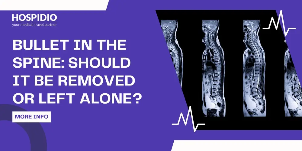
Few things on a scan feel as alarming as a bullet sitting beside the spinal cord. Most people's first instinct is simple: take it out. Yet many patients are told by their surgeons that the bullet should stay where it is, and families are left wondering whether that advice reflects caution, limited resources or genuine medical judgement.
The honest answer is that it depends, and it depends on a small number of factors that can be explained clearly. In this guide we walk through each of them: where the bullet lies, what it is doing to the nerves, whether it threatens infection or lead exposure, and what removal can and cannot achieve. Whether the injury happened last month or ten years ago, this is the reasoning a spine surgeon will apply to your scans.
Rapido verdetto
Removal is most likely to help when the bullet lies inside the spinal canal at the lower end of the spine (around T12 and below), when the injury is incomplete, or when nerve function is getting worse.
Removal is unlikely to restore function after a complete injury in the chest region of the spine, and it carries real risks.
Bullets in muscle or embedded in bone beside the spine are usually left alone.
Clear reasons to remove a bullet at any stage include infection, a persistent spinal fluid leak, a bullet that is moving and lead poisoning.
New weakness or numbness years after the injury needs prompt assessment. The cause is not always the bullet.
Where the Bullet Is Matters More Than the Fact That It Is There
"Near the spine" covers very different situations. A bullet in the back muscles and a bullet inside the spinal canal are entirely different problems. A thin-slice CT scan shows exactly where it lies, and that location drives most of the decision.
| Where the Bullet Lies | Cosa significa di solito | Approccio usuale |
| Muscle or soft tissue beside the spine | Sealed off by scar tissue; no pressure on nerves | Leave alone unless painful, infected or close to the skin |
| Embedded in a vertebra (the bone) | Bone usually heals around it | Usually leave alone |
| In the disc space between vertebrae | Can wear on the disc; rare reports of lead absorption | Assess individually; monitor blood lead |
| In a facet joint (the small joints at the back of the spine) | Joint fluid can dissolve lead and damage the joint | Removal often considered |
| Inside the spinal canal, outside the protective membrane | May press on the cord or nerve roots | Depends on level, completeness and symptoms |
| Inside the membrane, floating in spinal fluid | Can migrate; contact with fluid can release lead | Removal more often considered |
Dimension 1: Neurological Recovery
This is the question most families care about: will removing the bullet help them move again?
The best-known study on the subject followed 90 patients with bullet fragments in the spinal canal. Where the injury lay between the T12 and L4 levels, the lower part of the spine where the cord ends and the cauda equina nerve roots begin, patients who had the bullet removed recovered significantly more motor function than those who did not. In the chest region of the spine, from T1 to T11, removal made no meaningful difference to motor recovery.
There is a sound reason for this. Nerve roots in the lower spine behave more like the nerves in the arms and legs: they can recover if pressure is taken off them. The spinal cord itself, especially in the narrow thoracic canal, is usually damaged at the moment of impact, and taking out the bullet afterwards does not reverse that damage.
Two other factors shape the decision:
- Completeness of the injury. If some movement or sensation remains below the injury (an incomplete injury), there is something to protect, and relieving pressure is more likely to be worthwhile.
- Change over time. Nerve function that is getting worse, at any level, is a reason for urgent decompression.
In the neck, removing fragments from the canal may be justified in selected incomplete injuries, but each case needs careful individual judgement.
About Steroids: High-dose steroids were once given routinely after spinal cord injury. For gunshot injuries to the spine, a review of 254 patients found no neurological benefit and more complications in those who received them. They are no longer recommended for these injuries.
Dimension 2: Spinal Stability
Many patients assume a bullet in the spine means the spine is broken and needs screws and rods. In most civilian gunshot injuries, it does not. A bullet usually damages a small area of bone rather than collapsing the whole column. A large single-centre review of 159 patients concluded that most civilian spinal gunshot wounds can be treated without surgery, and that leaving them unoperated did not lead to late instability.
High-velocity injuries and blast injuries are different. They can shatter several vertebrae, and some do need fixation. The point is that fusion should be offered because a scan shows instability, not simply because a bullet is present.
Dimension 3: Infection and Spinal Fluid Leaks
Two complications can turn a "leave it alone" decision into a "take it out" decision.
- Infection: A bullet that passed through the bowel before reaching the spine can carry bacteria with it. Doctors usually treat this with a course of antibiotics, typically at least seven days, rather than routine removal. In the study above, about a fifth of the bullets had passed through the digestive tract and no spinal infections were recorded. A spinal abscess, a persistent fever, a draining wound over the spine or rising infection markers is a reason to operate.
- Spinal fluid leak: If the protective membrane around the cord (the dura) was torn and fluid keeps leaking, through the wound or into the tissues, there is a risk of meningitis. A persistent leak is one of the clearest reasons for surgery, and the bullet is usually removed during the repair.
Dimension 4: Lead Exposure
Most bullets contain lead. When lead sits in muscle or bone, it is usually walled off by scar tissue and stays inert. Problems arise when it is bathed in fluid that can slowly dissolve it.
In the spine, this means a bullet in a facet joint, in the disc space or floating in spinal fluid. Lead poisoning from bullets in these locations has been reported, though it is rare. Symptoms are vague and can appear years later: tiredness, abdominal pain, headaches, memory or mood changes and anaemia. A blood lead test is simple, and for bullets in these locations it is worth checking periodically. A rising lead level is a clear reason to remove the bullet.
Dimension 5: Pain
People often hope that removing the bullet will relieve their pain. The evidence is discouraging. In the study above, bullet removal did not significantly reduce pain or improve sensation.
That is because most pain after a spinal gunshot injury is nerve pain from the original damage to the cord or nerve roots, not from the metal itself. The exception is a bullet clearly pressing on a single nerve root and causing pain along that nerve, which may improve with removal. For most people, a dedicated pain specialist offers more relief than surgery.
Dimension 6: Imaging and MRI
A retained bullet affects future scans in two ways. First, metal creates artefact, a starburst of distortion that can hide the very area the doctor needs to see. Second, many patients are refused MRI altogether because of the bullet.
Most bullets are made of lead and copper, which are not magnetic, and radiologists can often judge from CT appearances whether a fragment is likely to be safe. Fragments close to the spinal cord still call for extra caution. When a bullet is not removed, alternatives such as CT myelography (a CT scan with dye injected into the spinal fluid) can show the cord and nerve roots instead of MRI.
Dimension 7: The Risks of Surgery Itself
Removing a bullet from the spine is a precise operation, but it is not risk-free. Possible complications include:
- spinal fluid leak from opening the membrane
- infezione
- further injury to the cord or nerve roots
- emorragia
- scar tissue that later tethers the cord
- instability if a large amount of bone must be removed, sometimes requiring fixation
These risks are why surgeons do not remove bullets simply because they are there. The benefit has to outweigh them.
Every bullet and every injury is different. Our partner spine neurosurgeons can review your scans and tell you, in writing, whether removal is likely to help before you make any travel plans.
Months or Years Later: When a Late Operation Makes Sense
Many people reading this were injured long ago, sometimes in another country, and are asking whether something should be done now. Late removal is reasonable when there is a specific, current problem:
- new or worsening weakness, numbness or bladder change
- a bullet that has moved on serial scans ??? infection, an abscess or a sinus over the spine
- a persistent spinal fluid leak or fluid collection
- rising blood lead levels
- a bullet in the lower canal clearly compressing nerve roots, with symptoms that match
What late removal rarely achieves is the recovery of function lost at the time of injury, especially after a complete injury higher in the spine.
New Weakness Years Later Is a Warning Sign
If someone with an old spinal gunshot injury starts losing strength or sensation, the bullet is not the only possible cause. A fluid-filled cavity can form in the cord (post-traumatic syringomyelia), or scar tissue can tether it. Both can cause slowly rising weakness months or years later, and both are treatable. This needs prompt imaging and review by a spine neurosurgeon.
What the Evidence Says About Outcomes
- We avoid quoting a single success rate for this surgery, because outcomes depend far more on the injury than on the operation. What the research does allow us to say:
- The completeness of the injury at the start is the strongest predictor of recovery. Incomplete injuries recover far more often than complete ones.
- Injuries in the thoracic spine are the most likely to be complete. In the 159-patient review, about a third of patients improved by at least one grade on the ASIA scale over follow-up.
- Removal of bullets in the lower canal (T12 to L4) is associated with better motor recovery. Removal higher in the spine is not.
- Surgery did not improve neurological outcomes overall in that review, but it remained the right treatment for infection and persistent spinal fluid leaks.
Be wary of any clinic that promises a specific chance of walking after bullet removal without first examining you and reviewing your scans.
Guida alla decisione
| tua situazione | Cosa succede di solito |
| Bullet in muscle or bone beside the spine, no symptoms from it | Leave alone; routine follow-up |
| Bullet in the canal at T12 or below, incomplete injury | Removal often recommended |
| Bullet in the canal in the chest region, complete injury, stable | Usually left alone; focus on rehabilitation |
| Neurological function getting worse at any level | Urgent imaging and decompression |
| Persistent spinal fluid leak, abscess or draining wound | Surgery to clear infection or repair the leak, usually with removal |
| Bullet in a facet joint, disc space or floating in spinal fluid | Assess for removal; check blood lead |
| Bullet has moved on serial scans | Removal often considered |
| Old injury, new weakness years later | Imaging for syrinx, tethering or migration before deciding |
| Pain is the main problem | Pain specialist first; surgery only if a clear nerve root cause |
Come funziona la valutazione
A sound recommendation rests on a few key investigations. Before you travel, we will usually ask for:
A thin-slice CT scan of the spine as original DICOM files, showing exactly where the bullet lies and whether the bone is stable
- Any MRI or CT myelogram already done
- Your neurological examination, ideally an ASIA assessment, with dates so change over time can be seen
- Operative notes and discharge summaries from the time of injury
- Details of any infection, wound discharge or spinal fluid leak, with culture reports
- Bladder and bowel function, and urodynamic studies if available
- Blood tests, including a blood lead level if the bullet is in a joint, disc or spinal fluid
Send the Scan Files, Not Photos: A phone photograph of a CT screen cannot show the bullet's exact relationship to the cord and nerve roots. Ask the hospital that did your scan for the DICOM files on a disc or by secure download.
Treatment in India for International Patients
Spine neurosurgery teams at India's major tertiary hospitals manage penetrating spinal injuries alongside the wider care these patients need: neurology, infectious disease, urology and spinal cord injury rehabilitation. For international patients, a typical pathway looks like this:
- Remote review: Your scans and reports are reviewed before travel, and you receive a written opinion on whether surgery is recommended.
- Arrival and assessment: Repeat examination and any further imaging, usually within the first few days.
- Surgery, if advised: Removal with or without decompression or repair of the membrane, and fixation only if the spine is unstable.
- Hospital stay: Usually 5 to 10 days after surgery, longer if infection or a spinal fluid leak needs treatment.
- Rehabilitation: Inpatient rehabilitation in India, or a home programme with remote follow-up.
- Total time in India: Typically 2 to 3 weeks for removal alone, and longer when rehabilitation is added.
Patients from eligible countries can apply online for an e-Medical Visa, with up to two companions on e-Medical Attendant Visas. Rules vary by nationality, so check the official Indian visa portal before booking.
Cost of Bullet Removal From the Spine in India
The ranges below are indicative for private tertiary hospitals in India and are intended for planning only. A firm estimate follows review of your scans.
| Trattamento | Costo indicativo in India (USD) |
| Specialist review of scans and reports (remote) | Free for written opinion$10 to $30 for video consultation |
| Bullet removal with decompression (laminectomy), no fixation | 6000 $ a $ 8500 |
| Bullet removal with spinal fixation | 7500 $ a $ 11500 |
| Repair of a persistent spinal fluid leak | 5000 $ a $ 8000 |
| Surgery for spinal infection or abscess | 6000 $ a $ 10000 |
| Surgery for post-traumatic syrinx or cord tethering | 9000 $ a $ 11000 |
| Riabilitazione per pazienti ricoverati con lesioni del midollo spinale, al mese | 4000 $ a $ 7000 |
Figures exclude flights, accommodation and food. Costs rise when infection, prolonged intensive care or multiple-level fixation is involved.
Leggi anche: Gunshot Blast Injury Treatment in India
Blog recenti


Domande frequenti
Often, yes. Bullets in the muscles or bone beside the spine are usually sealed off by scar tissue and cause no further harm. Bullets inside the spinal canal need individual assessment, as removal can help in some situations, particularly in the lower spine. Leaving a bullet is safest when it is stable and not causing infection, leaks or worsening symptoms. Your surgeon will usually recommend periodic follow-up, and a blood lead test if the bullet sits in a joint, the disc space or spinal fluid.
Rarely, but it can. A bullet floating inside the membrane around the spinal cord can shift with posture and gravity, occasionally causing new symptoms such as leg pain, weakness or bladder changes. Bullets embedded in bone or sealed in muscle almost never move. Comparing serial CT scans or X-rays shows whether a bullet has changed position over time. A bullet that has moved and is causing symptoms is one of the recognised reasons to consider removal, even years after the injury.
It depends on the level and completeness of the injury. Removal from the lower spinal canal, around T12 and below, is associated with better motor recovery, especially in incomplete injuries. After a complete injury in the chest region of the spine, removing the bullet rarely restores walking, because the cord was damaged at the moment of impact. A specialist examination and a review of your scans give the most realistic picture. Rehabilitation often contributes more to independence than surgery does.
It can, but it is uncommon. Lead poisoning from a bullet usually happens when the lead is bathed in fluid that slowly dissolves it, such as joint fluid in a facet joint, the disc space or spinal fluid. Bullets sealed in muscle or bone rarely release meaningful lead. A simple blood test measures your lead level. Symptoms can include tiredness, abdominal pain, headaches, memory or mood changes and anaemia, and may appear years after the injury.
Sometimes. Most bullets are made of lead and copper, which are not magnetic, and radiologists can often judge the likely composition from CT scans. Because the spinal cord is so close, the decision needs a formal safety assessment. The metal can also distort MRI images, so CT myelography is sometimes used instead. Bring your CT scan and any information about the injury. Avoid any scan where staff have not been told about the bullet.
New weakness years later can come from a fluid-filled cavity forming in the spinal cord (post-traumatic syringomyelia), scar tissue tethering the cord, a bullet that has moved, or infection. These causes are treatable, but they need prompt imaging and review by a spine neurosurgeon. Do not assume that the change is simply part of the old injury. Keep a record of when the change started and what has changed, including bladder and bowel function, to help the specialist.
Usually not. Research found that removing bullets from the spinal canal did not significantly reduce pain, because most pain after these injuries is nerve pain from the original damage, not from the metal. The exception is a bullet clearly pressing on a single nerve root and causing pain along that nerve, which may improve after removal. A pain specialist can offer medicines, nerve blocks and other treatments designed specifically for nerve pain.
Usually not. Most civilian gunshot injuries damage a small area of bone and leave the spine stable, and studies have found that treating them without surgery does not lead to later instability. Fusion is needed when scans show the spine is unstable, more often after high-velocity or blast injuries, or when surgery removes a large amount of bone. Your surgeon should explain, from the CT scan, why fixation is or is not needed in your case.
Most patients stay about two to three weeks in total for removal alone, including assessment, surgery and a hospital stay of roughly five to ten days. Treatment for infection or a spinal fluid leak, or a period of inpatient rehabilitation, extends this. Your written plan will give an expected length of stay before you travel. Many follow-up reviews can be done remotely once you return home.
Send your CT scans as DICOM files, any MRI, operative notes and recent neurological assessments to [email protected] or by WhatsApp to +91-9319955321. We arrange a review by a spine neurosurgeon and send you a written opinion on whether removal is advisable, the expected stay and an estimated cost. The review is free and there is no obligation to travel. If surgery is unlikely to help, we will tell you.
Guneet Bhatia è la fondatrice di HOSPIDIO e un'esperta revisore di contenuti con una vasta esperienza nello sviluppo di contenuti medici, nella progettazione didattica e nel blogging. Appassionata della creazione di contenuti di grande impatto, eccelle nel garantire accuratezza e chiarezza in ogni articolo. Guneet ama intavolare conversazioni significative con persone di diverse origini etniche e culturali, arricchendo così la sua prospettiva. Nel tempo libero, si dedica alla famiglia, ascolta buona musica e si diverte a ideare soluzioni innovative con il suo team.
Dr. Hamza Shaikh è un chirurgo vertebrale di grande esperienza, con oltre 15 anni di attività nel settore, specializzato nella gestione completa di deformità spinali come scoliosi e cifosi, nonché di patologie spinali complesse. Attualmente ricopre il ruolo di chirurgo vertebrale consulente presso il Manipal Hospital di Dwarka, dove fornisce cure spinali avanzate e basate sull'evidenza scientifica, personalizzate sia per pazienti pediatrici che adulti.