
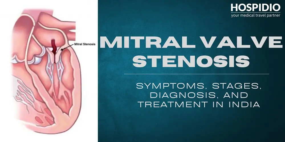
Mitral valve stenosis, also known as mitral stenosis, is a heart valve condition in which the mitral valve opening becomes narrower than normal. As a result, it cannot allow enough blood to flow from the left atrium (the upper chamber of the heart) into the left ventricle (the lower chamber). The left ventricle is responsible for pumping oxygen-rich blood throughout the body.
This narrowing can increase pressure inside the heart and lungs, potentially causing symptoms such as shortness of breath, fatigue, and heart palpitations. Understanding mitral valve stenosis symptoms, diagnosis, and treatment options can help patients make informed decisions about their heart health.
Mitral valve stenosis is relatively uncommon in developed countries but remains more prevalent in developing regions, particularly where access to antibiotics and medical care is limited. The condition is also more common in females.
¿Qué es la estenosis de la válvula mitral?
The mitral valve is located between the left atrium and the left ventricle. It opens to let oxygen-rich blood pass into the left ventricle and closes to prevent blood from flowing backward.
When the valve becomes thickened, stiff, or fused, its opening narrows. This makes it harder for blood to move through the heart, increasing pressure in the left atrium and potentially in the blood vessels of the lungs.
Mitral valve problems can affect how the valve opens or closes:
- Estenosis mitral: The valve does not open fully, restricting forward blood flow.
- Prolapso de la válvula mitral: The valve leaflets bulge backward into the left atrium when the heart contracts. This can sometimes cause leakage.
- Regurgitación de la válvula mitral: The valve does not close properly, allowing blood to flow backward into the left atrium.
These conditions are different, although more than one valve problem can occur in the same patient.
¿Qué causa la estenosis de la válvula mitral?
Las causas comunes incluyen:
- Fiebre reumática: An inflammatory complication of certain untreated streptococcal infections can damage the valve and cause scarring years later.
- Calcificación relacionada con la edad: Calcium deposits stiffen the valve.
- Defectos cardíacos congénitos: Some people are born with a narrowed or abnormally formed valve.
Most cases are acquired rather than directly inherited, although some congenital defects may have genetic links.
What are the Symptoms of Mitral Valve Stenosis?
Mild mitral valve stenosis may not cause noticeable symptoms for years. As the narrowing becomes more severe, symptoms may develop or worsen.
Los síntomas comunes incluyen:
- Shortness of breath during activity or lying down.
- Fatigue and reduced stamina.
- Palpitaciones o latidos cardíacos irregulares.
- Tos persistente, a veces con sangre.
- Swelling in the feet, ankles, or abdomen.
- Infecciones respiratorias frecuentes.
- Dizziness and difficulty with daily activities.
Some people first notice symptoms during pregnancy or strenuous physical activity, when the heart needs to pump more blood. Symptoms such as sudden severe breathlessness, fainting, or coughing up significant amounts of blood require urgent medical assessment.
How is Mitral Valve Stenosis Diagnosed?
A cardiologist evaluates your symptoms, medical history, physical examination findings, and diagnostic test results to confirm mitral valve stenosis and determine its severity.
| Prueba de diagnóstico | Qué evalúa |
| Ecocardiograma. | Uses ultrasound to assess valve structure, blood flow, pressure gradients, and the severity of narrowing. |
| Ecocardiograma transesofágico (ETE) | Provides detailed images of the valve and may help identify blood clots before a procedure. |
| Electrocardiograma (ECG / EKG) | Detects abnormal heart rhythms, including atrial fibrillation. |
| Radiografía del pecho. | May show enlargement of the heart or signs of fluid congestion in the lungs. |
| TC o RM cardiaca | Provides additional structural information when needed. |
| Cateterización cardiaca | Measures pressures within the heart and helps clarify findings in selected cases. |
Echocardiography is the primary test used to diagnose mitral valve stenosis. Your cardiologist may recommend additional investigations depending on the initial findings and the treatment being considered.
If you've been diagnosed with mitral valve stenosis and are unsure about the next steps, our patient consultant can help you explore treatment options in India. Talk to Our Patient Consultant
What Complications Can Mitral Valve Stenosis Cause?
Without appropriate monitoring and treatment, significant mitral valve stenosis can lead to:
- Fibrilación auricular: An irregular heart rhythm that can increase the risk of blood clots.
- Carrera: A blood clot may travel to the brain and interrupt its blood supply.
- Hipertensión pulmonar: Increased pressure in the blood vessels supplying the lungs.
- Insuficiencia cardiaca: The heart may struggle to maintain effective circulation.
- Edema pulmonar: Fluid accumulation in the lungs that can cause severe breathing difficulties.
- Coágulos de sangre: Reduced blood flow and abnormal heart rhythms can promote clot formation.
- Reduced physical capacity: Fatigue and breathlessness may interfere with everyday activities.
Regular cardiac assessments can help detect progression and guide timely treatment.
Mitral Valve Stenosis During Pregnancy
Pregnancy increases blood volume and the workload on the heart. For women with mitral valve stenosis, particularly moderate or severe disease, these changes can worsen breathlessness and increase the risk of abnormal heart rhythms, pulmonary hypertension, and heart failure.
Women with known mitral stenosis should ideally undergo a cardiac assessment before becoming pregnant. During pregnancy, care may involve a cardiologist, a high-risk obstetrician, and other specialists.
Depending on the severity of the condition, management may include:
- Regular monitoring of heart function and symptoms.
- Carefully selected medications to control heart rate or fluid buildup.
- Individualized planning for labor, delivery, and postpartum care.
- Balloon mitral commissurotomy during pregnancy in selected patients whose symptoms remain severe despite medical treatment.
Open-heart surgery during pregnancy is generally reserved for situations in which maternal life is at risk and other suitable options are unavailable or unsuccessful.
If you are pregnant or planning a pregnancy and have mitral valve stenosis, consult a cardiologist before making decisions about medication or treatment. Never start or stop heart medication without medical advice.
What are the Treatment Options for Mitral Valve Stenosis?
Treatment depends on the severity of narrowing, symptoms, valve anatomy, and overall health. Some patients need regular monitoring, while others may require a procedure or surgery.
1. Medicación
Medications help manage symptoms and complications but cannot reverse valve narrowing.
- Diuréticos: Reducir la acumulación de líquido.
- Bloqueadores beta: Ayuda a controlar la frecuencia cardíaca.
- Anticoagulantes: Reduce blood clot risk when indicated.
- Medicamentos antiarrítmicos: Manage abnormal heart rhythms.
- antibióticos: Treat bacterial infections or prevent recurrent rheumatic fever when indicated.
2. Procedimientos basados en catéter
These minimally invasive procedures use a thin tube inserted through a blood vessel to reach the heart.
- Valvulotomía mitral con balón: Also called balloon mitral valvuloplasty, this procedure uses a balloon-tipped catheter to widen the narrowed valve opening by separating fused valve leaflets. It may be suitable for patients with rheumatic mitral stenosis, favorable valve anatomy, and no significant contraindications.
- Reemplazo de válvula mitral transcatéter: involves implanting a replacement valve through a catheter without conventional open-heart surgery. It is suitable only for selected patients, depending on their condition and valve anatomy.
3. Tratamiento quirúrgico
Surgery may be recommended when the valve is severely damaged, balloon treatment is unsuitable, or other valve abnormalities require correction.
Surgical Commissurotomy
Commissurotomy involves separating fused areas of the mitral valve to improve blood flow. It can be performed using two approaches:
- Closed mitral commissurotomy: The surgeon separates the fused valve commissures without directly visualizing the valve. This approach is now less commonly used.
- Open mitral commissurotomy: The surgeon directly visualizes the valve and separates the fused commissures, allowing a more detailed assessment of the valve structure. Conventional open surgery requires cardiopulmonary bypass.
The decision depends on factors such as age, expected durability, bleeding risk, pregnancy plans, and the ability to maintain anticoagulation monitoring.
Recovery After Treatment and Long Term Follow-Up
Recovery varies according to whether treatment involves medication, balloon valvotomy, or surgery.
After balloon valvotomy, patients may need a shorter hospital stay and can often resume normal activities sooner than after open-heart surgery. Recovery following surgical valve repair or replacement generally takes longer and may require several weeks or more.
Los cuidados a largo plazo pueden incluir:
- Follow up appointments with a cardiologist.
- Echocardiograms to assess valve function and heart pressures.
- Monitoring for abnormal heart rhythms and other complications.
- Anticoagulation monitoring when required.
- Cardiac rehabilitation and gradual return to activity when recommended.
- Review of symptoms, medications, and lifestyle measures.
Mitral Valve Stenosis Treatment Cost in India
The cost of mitral valve stenosis treatment in India depends on the procedure, hospital, specialist fees, valve type, and the patient's overall health. Treatment may involve medication, valvotomía mitral con balón, surgical commissurotomy, or reemplazo de la válvula mitral.
Compared with the US and other countries with higher healthcare costs, cardiac treatment in India can be significantly more affordable, while offering access to specialized cardiac care. Actual costs vary depending on the patient's condition and the recommended treatment.
HOSPIDIO can help international patients explore suitable hospitals, coordinate treatment arrangements, and obtain a personalized cost estimate based on their medical reports and clinical needs.
Los mejores cardiólogos de la India




Conclusión
Mitral valve stenosis can progress gradually, and some people remain symptom-free for years. However, significant narrowing can affect circulation, increase pressure in the lungs, and lead to serious complications if left untreated.
The right approach depends on accurate diagnosis and an individualized assessment of valve anatomy, symptoms, and overall health. While medication and regular monitoring may be sufficient for some patients, others may benefit from balloon valvotomy or surgical valve treatment.
If you or a loved one has been diagnosed with mitral valve stenosis and is considering treatment in India, HOSPIDIO can help you explore suitable hospitals, coordinate medical consultations, and arrange follow-up care throughout your treatment journey.
Blogs



Preguntas frecuentes
Focus on healthy food options and follow the diet plan recommended by your doctor or nutritionist. Your dietary requirements may vary depending on your recovery, overall health, and medications.
Avoid heavy lifting, strenuous exercise, and activities that strain your chest, especially after open-heart surgery. Follow your surgeon's advice on driving, returning to work, and resuming exercise. Gradually increase physical activity as recommended.
Watch for fever, chills, redness, swelling, warmth, or discharge around the surgical incision. Contact your healthcare team promptly if these symptoms develop. For cardiac care in India, HOSPIDIO can help you connect with suitable hospitals and specialists.
Conditions such as diabetes, high blood pressure, kidney disease, or lung disorders may affect surgery and recovery. Discuss your medical history with your cardiologist, or connect with HOSPIDIO to explore suitable cardiac care options in India.
Mechanical mitral valves can last for many years but generally require lifelong blood-thinning medication. Biological valves may wear out over time and require another intervention. Regular echocardiograms help monitor valve function.
Yes, mitral valve stenosis can recur after balloon valvotomy if the valve becomes narrowed again over time. Regular cardiology consultations and echocardiograms help detect recurrent narrowing and determine whether further treatment is necessary.
Shruti Verma se licenció en Biotecnología y cuenta con experiencia en redacción médica y desarrollo de contenido científico. Se especializa en traducir información biomédica y sanitaria compleja en contenido claro, preciso y accesible para diversos públicos. Cuando no está diseñando contenido, probablemente se dedica al diseño gráfico.
Sasmita Bal es especialista en marketing digital y creación de contenido en HOSPIDIO, con experiencia en SEO y contenido internacional sobre salud. Revisa el material publicado para asegurar su optimización para motores de búsqueda y su relevancia para las necesidades de los pacientes internacionales que buscan tratamiento en la India. Todo el contenido que revisa es redactado y aprobado clínicamente por el fundador de HOSPIDIO y los especialistas médicos pertinentes antes de su publicación.