
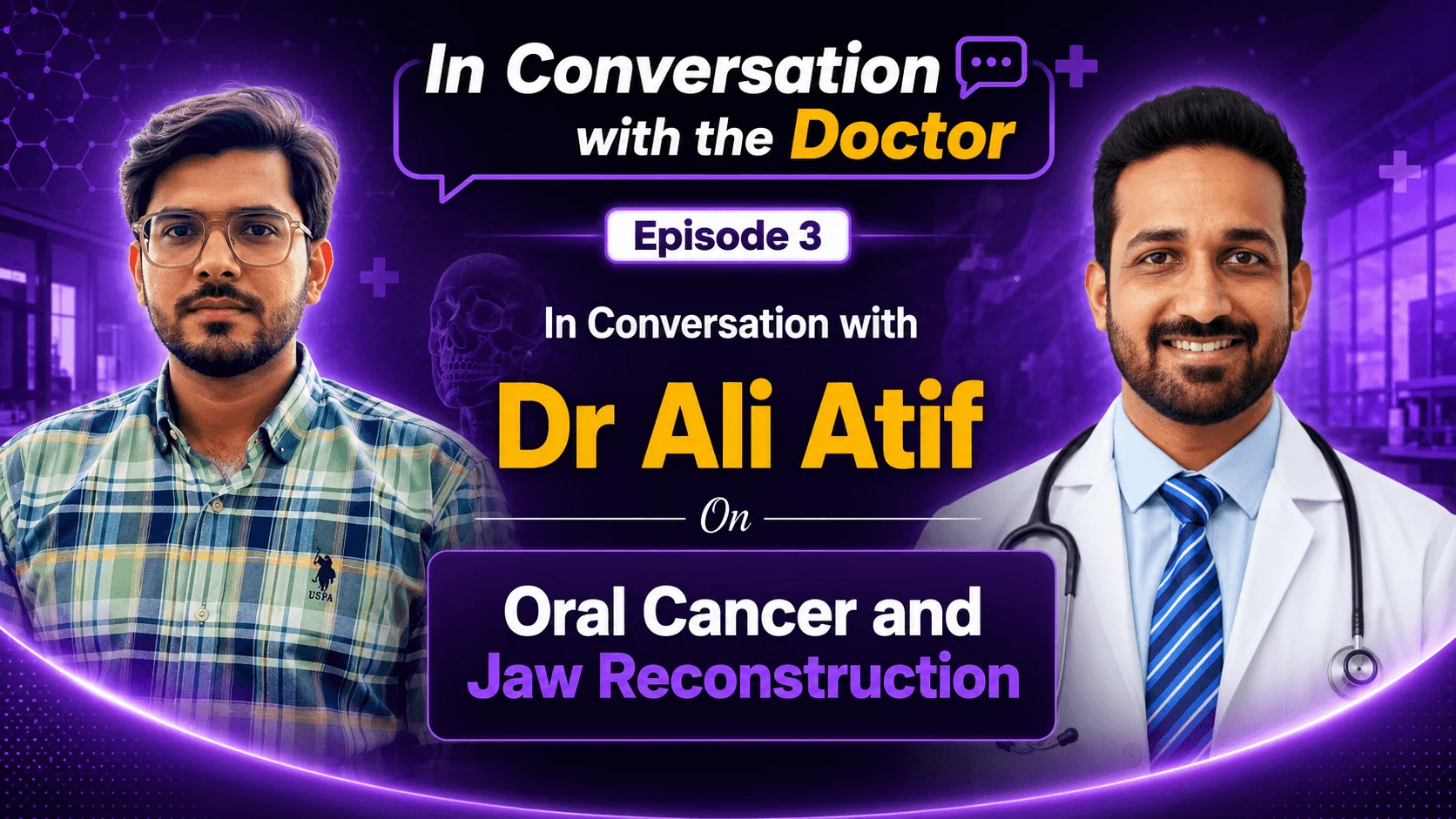
In questa sessione di domande e risposte, Dr. Ali Atif, chirurgo orale e maxillo-facciale di Lucknow con 15 anni di esperienza in oncologia cervico-facciale, risponde alle domande più frequenti che pazienti e familiari si pongono sul cancro orale, dai primi segnali di allarme e fattori di rischio alla chirurgia, alla ricostruzione della mandibola e alle prospettive future.
Guarda la conversazione completa:
D: Per chi non ha mai sentito parlare di "cancro orale", di cosa si tratta esattamente, dove può svilupparsi e perché è considerato così grave?
Il cancro orale ha origine all'interno della cavità orale, da cui il nome, ma tende a diffondersi anche in altre parti del corpo. Può svilupparsi sulla lingua, sul labbro, sul pavimento della bocca, sulla guancia o persino sulla mandibola.
In una persona sana, le cellule si sviluppano e si dividono in modo controllato e normale. In un paziente affetto da cancro, le cellule perdono questa normalità e iniziano a moltiplicarsi a un ritmo molto rapido e inarrestabile, formando una massa tumorale. Questa massa può diffondersi dalla cavità orale ai linfonodi e da lì ai polmoni, alle ossa e ad altri organi distanti. I tumori del cavo orale sono particolarmente aggressivi rispetto a molti altri tumori, ed è proprio per questo che la diagnosi precoce e la consapevolezza sono così importanti.
D: Chi è maggiormente a rischio di sviluppare un tumore della bocca? Tabacco, alcol e noce di betel sono spesso citati come fattori di rischio principali, qual è il fattore determinante più importante?
Il consumo di tabacco è di gran lunga il principale fattore di rischio. Questo include il tabacco da masticare, molto diffuso in India, e il tabacco senza fumo, ampiamente utilizzato nei paesi africani.
L'uso di alcol insieme al tabacco ha un effetto sinergico, aumentando ulteriormente il rischio. La masticazione di noce di areca o di betel può portare a una condizione chiamata fibrosi sottomucosa orale, in cui si sviluppa fibrosi intorno alla bocca e si riduce l'apertura della bocca nel tempo, una condizione precancerosa che può successivamente evolvere in cancro. L'infezione da papillomavirus umano (HPV) è un altro fattore contribuente; le biopsie di lesioni cancerose rimosse a volte mostrano la presenza di HPV. Tra tutti questi fattori, il tabacco rimane il principale responsabile.
D: Quali sono i primi segnali di allarme del cancro orale che i pazienti spesso ignorano o scambiano per qualcosa di innocuo, come un'ulcera? Quanto tempo aspettano in genere i pazienti prima di consultare un medico e perché questo ritardo è importante?
Non tutte le ulcere della bocca sono tumori orali, quindi non c'è motivo di allarmarsi per ogni lesione. Detto questo, alcuni segnali meritano attenzione: un'ulcera che persiste per molti mesi, macchie bianche all'interno della bocca, fibrosi che riduce gradualmente l'apertura della bocca, una sensazione di bruciore quando si mangiano cibi piccanti o un nodulo duro in bocca o sul collo.
La maggior parte dei pazienti non pensa subito che possa trattarsi di cancro. In genere, si rivolgono prima al dentista per quella che sembra una semplice ulcera, ed è proprio per questo che un dentista specializzato in oncologia orale è spesso la figura più indicata per una diagnosi precoce. La trasformazione da una semplice ulcera a un tumore del cavo orale non avviene in un solo giorno. Il corpo di solito manifesta prima segni precancerosi, come la leucoplachia, una macchia bianca all'interno della guancia che non si può rimuovere con un raschietto, o una progressiva riduzione dell'apertura della bocca. Se un sintomo persiste e appare sospetto, è consigliabile consultare un medico o un dentista senza indugio.
D: Potrebbe spiegarci cos'è un tumore alla mandibola, dove si sviluppa e in cosa si differenzia dai tumori del cavo orale che colpiscono i tessuti molli come la lingua e la guancia?
È fondamentale distinguere innanzitutto un tumore, ovvero una patologia maligna, da un tumore, che in genere è una crescita benigna e autolimitante. Un tumore richiede un intervento chirurgico completo, inclusa la dissezione del collo per rimuovere i linfonodi infetti, seguita da un intervento di ricostruzione. Un tumore, invece, è una crescita circoscritta che comporta un certo rischio di evolvere in tumore, ma non è di per sé un tumore.
I tumori della mascella spesso hanno origine da problemi dentali; ad esempio, un dente infetto non trattato può trasformarsi in una cisti, che nel tempo può evolvere in un tumore. L'ameloblastoma ne è un esempio ben noto. Alcuni tumori crescono verso l'esterno, espandendosi e gonfiando visibilmente la mascella, mentre altri crescono verso l'interno e passano inosservati finché un dente non si allenta o il gonfiore non diventa evidente. Se trattati in modo appropriato, questi tumori sono autolimitanti e possono essere rimossi senza metastatizzare. Le visite dentistiche di controllo di routine sono importanti proprio per individuarli precocemente.
Per saperne di più: Trattamento del cancro orale in India
D: L'ameloblastoma è uno dei tumori della mascella più discussi. Perché desta tanta preoccupazione nonostante sia benigno, e cosa succede se non viene trattato?
L'ameloblastoma è un tumore raro a crescita lenta che di solito si forma nella mandibola, nella regione dei denti posteriori, vicino alla posizione tipica dei denti del giudizio. Si espande internamente, riassorbendo l'osso circostante man mano che cresce, e può diffondersi ampiamente in più direzioni.
Ciò che lo rende particolarmente preoccupante è il suo potenziale maligno. Se non trattato, può evolvere in cancro nel tempo. Per questo motivo, la diagnosi inizia sempre con una biopsia per escludere la presenza di un tumore, seguita da un piano di trattamento ricostruttivo definitivo. Poiché cresce molto lentamente e spesso passa inosservato, una diagnosi tardiva a volte significa dover rimuovere una porzione significativa, o addirittura tutta, la mandibola.
D: Come si manifesta tipicamente un ameloblastoma? In quale fase iniziano a comparire sintomi come alitosi o gonfiore?
A differenza di molte altre patologie, nelle fasi iniziali si tratta perlopiù di un gonfiore indolore, motivo per cui spesso i pazienti non si rivolgono subito al medico. Quando inizia a manifestarsi il dolore, di solito è segno che il tumore sta iniziando a coinvolgere i denti, causandone l'allentamento. Questo crea degli spazi in cui il cibo può incastrarsi e può insorgere un'infezione in prossimità del tumore stesso, con conseguente alitosi e gengive rosse e gonfie.
Questa è in genere la fase in cui i pazienti finalmente cercano cure, più tardi rispetto all'ideale, ma anche a questo punto la condizione rimane curabile se non viene ulteriormente ignorata.
D: Potrebbe descriverci in dettaglio cosa succede durante un intervento chirurgico per il cancro orale? Cosa viene rimosso, quanto sono ampi i margini chirurgici e come fanno i chirurghi ad assicurarsi che tutto il tumore venga asportato?
I tumori del cavo orale vengono classificati secondo il sistema TNM, che tiene conto delle dimensioni del tumore, del numero di linfonodi coinvolti e del potenziale di metastasi, ovvero della presenza di metastasi in linfonodi distanti. I chirurghi valutano questo aspetto tramite un esame obiettivo, palpando la zona interessata alla ricerca di margini induriti e ispessiti, che indicano la presenza di tessuto canceroso.
Durante l'asportazione del tumore, si preleva un margine di sicurezza di circa 2.5 cm di tessuto sano circostante, anche se il tumore visibile appare piccolo, per ridurre al minimo il rischio di recidiva. Si tratta necessariamente di un intervento chirurgico aggressivo e ablativo, poiché margini eccessivamente conservativi aumentano significativamente la probabilità di ricomparsa del tumore.
Oltre alla rimozione del tumore primario, viene eseguita una dissezione del collo per asportare i linfonodi a vari livelli (dal livello 1 al livello 5) che potrebbero essere infetti. Questi vengono inviati per la biopsia al fine di determinare con precisione quali livelli siano interessati, il che a sua volta guiderà le decisioni in merito alla necessità di radioterapia, chemioterapia o entrambe.
D: Se una parte della mandibola di un paziente deve essere rimossa, cosa succede dopo? La ricostruzione mandibolare può davvero restituire una vita normale?
Sì. La chirurgia ricostruttiva moderna ha fatto molta strada. Grazie alla modellazione digitale e alle strutture mandibolari stampate in 3D, i chirurghi possono ora ricreare una porzione mancante della mandibola con un elevato grado di precisione. Una delle tecniche più consolidate è l'innesto libero di perone, in cui un osso della gamba del paziente (il perone) viene prelevato, modellato in una struttura simile a una mandibola e posizionato nella regione interessata completo del proprio apporto sanguigno.
In seguito, nell'osso ricostruito possono essere inseriti anche impianti dentali, consentendo ai pazienti di tornare a una vita sostanzialmente normale, compresa la capacità di mangiare e parlare, dopo il processo di guarigione.
D: Oltre al trapianto di perone libero, quali altre opzioni di ricostruzione sono disponibili?
Per la ricostruzione della guancia o dei tessuti molli, si ricorre spesso a lembi localizzati quando un lembo libero non è economicamente vantaggioso o clinicamente indicato. Le opzioni includono il lembo nasolabiale (che utilizza la cute nasale per ricostruire il pavimento della bocca), i lembi muscolari del collo e il lembo del muscolo grande pettorale, che utilizza il muscolo del torace per ricostruire il rivestimento interno della guancia o per ottenere una ricostruzione più completa della guancia.
Nei casi più complessi, in particolare nella ricostruzione della lingua dopo un'emiglossectomia (asportazione parziale della lingua), si può utilizzare un lembo libero radiale dell'avambraccio. Sorprendentemente, i pazienti sottoposti a questo tipo di ricostruzione spesso riescono a recuperare una capacità di parola quasi normale, a volte al punto che è difficile accorgersi che hanno subito un intervento chirurgico importante.
D: Quali sono i risultati funzionali ed estetici realistici dopo la ricostruzione?
I risultati sono generalmente molto buoni, sebbene possano verificarsi un certo indurimento o la formazione di cicatrici, soprattutto nei pazienti sottoposti anche a radioterapia o chemioterapia, entrambe in grado di influenzare la guarigione dei tessuti. Spesso, in seguito, si ricorre a trattamenti laser e altri interventi estetici per correggere le cicatrici e migliorare la simmetria.
Grazie alla modellazione digitale, i chirurghi possono creare un modello 3D del cranio del paziente, adattare la ricostruzione al lato controlaterale sano e ottenere risultati spesso indistinguibili da quelli di un paziente che non ha subito un intervento chirurgico importante.
D: Quanto dura un intervento chirurgico di resezione e ricostruzione importante della mandibola e cosa sperimenta il paziente nei giorni e nelle settimane successive?
La durata dell'intervento dipende in larga misura dal metodo di ricostruzione utilizzato. La rimozione del tumore primario e la dissezione del collo richiedono in genere dalle 2 alle 2.5 ore. L'aggiunta di un lembo locale comporta un'ulteriore 1 o 2 ore, portando la durata totale a circa 3 o 4 ore per l'intervento con ricostruzione mediante lembo locale.
La ricostruzione con lembo libero richiede tempi considerevolmente più lunghi, poiché prevede il collegamento dei vasi sanguigni tra il sito donatore e la cavità orale. La ricostruzione con lembo libero a base ossea può richiedere dalle 7 alle 8 ore o più nei casi complessi, mentre i lembi liberi di tessuto molle, come il lembo radiale dell'avambraccio, richiedono in genere dalle 5 alle 6 ore.
Subito dopo l'intervento, i pazienti vengono tenuti in terapia intensiva per il primo giorno, spesso con un sondino nasogastrico per l'alimentazione e talvolta con una tracheostomia per proteggere le vie aeree. Entro il secondo o terzo giorno, i pazienti vengono incoraggiati a camminare e a recuperare la mobilità. Poiché l'interno della bocca è in fase di guarigione, l'alimentazione avviene esclusivamente tramite sondino nasogastrico per circa 7-10 giorni, dopodiché vengono rimossi i punti di sutura e il paziente può iniziare a mangiare cibi morbidi o liquidi per via orale. La guarigione completa richiede in genere circa 21 giorni, momento in cui sono disponibili i risultati della biopsia e la stadiazione per guidare eventuali trattamenti successivi, come la radioterapia o la chemioterapia. La maggior parte dei pazienti viene dimessa intorno al terzo giorno post-operatorio, poiché le prime 72 ore rappresentano il periodo più critico.
D: Qual è la cosa più efficace che una persona può fare per ridurre il rischio di cancro orale?
Evitare tabacco, alcol e altre sostanze è il passo più importante in assoluto. Anche le pratiche sessuali sicure sono fondamentali, dato il ruolo che l'HPV può svolgere. Una dieta sana ed equilibrata, ricca di verdura, antiossidanti e proteine, contribuisce a ridurre il rischio complessivo, soprattutto per chi soffre di patologie concomitanti come diabete o ipertensione, oltre ad altri fattori di rischio.
Le visite dentistiche di routine sono uno strumento di prevenzione sottovalutato ma estremamente efficace. Chiunque smetta di fumare dopo aver appreso di questi rischi dovrebbe anche prendere l'abitudine di rimanere in contatto regolarmente con uno studio dentistico specializzato in oncologia orale, poiché questi specialisti sono nella posizione migliore per individuare i problemi nella fase iniziale, quando sono più facilmente curabili.
D: Cosa la entusiasma di più riguardo al futuro del trattamento del cancro orale e dei tumori della mascella, della chirurgia robotica, delle terapie mirate, di materiali ricostruttivi migliori o dell'intelligenza artificiale nella pianificazione chirurgica?
Tutte le risposte precedenti. La robotica ha già avuto un impatto significativo sui risultati chirurgici. I vaccini contro l'HPV e la ricerca in corso sulla vaccinazione contro il cancro orale rappresentano sviluppi promettenti sul fronte della prevenzione. La ricerca sulle cellule staminali sta aprendo la strada alla generazione di tessuti e scaffold più sani, riducendo potenzialmente la necessità di interventi chirurgici altamente ablativi e minimizzando la morbilità.
Le terapie mirate, la pianificazione chirurgica assistita dall'intelligenza artificiale e i continui finanziamenti globali per la ricerca stanno spingendo il settore verso trattamenti meno invasivi e più precisi, con conseguenti migliori risultati in termini di qualità della vita per i pazienti.
D: Per i pazienti in Africa o in Medio Oriente, dove il trattamento del cancro orale e dei tumori della mascella potrebbe non essere disponibile o troppo costoso, recarsi in India è un'opzione realistica? Cosa dovrebbero valutare nella scelta di un centro?
Sì, e sempre più spesso. Molti pazienti provenienti da queste regioni si trovano ad affrontare la mancanza di infrastrutture specializzate o costi semplicemente inaccessibili, anche laddove siano presenti medici qualificati in loco. L'India è diventata una destinazione importante per questo tipo di cure, con sistemi ben consolidati per la pianificazione di ogni fase del viaggio, dal prelievo in aeroporto e l'alloggio alla dieta e alle cure di follow-up.
L'India ospita un gran numero di oncologi di grande esperienza e gli ospedali locali hanno curato un numero significativo di pazienti provenienti dall'Africa e dal Medio Oriente, potendo contare su solide reti di riferimento. Sia l'accesso alle competenze mediche che la logistica per recarsi a ricevere le cure sono diventati considerevolmente più accessibili ed economici negli ultimi anni.
Chi siamo Dr. Ali Atif
Dr. Ali Atif è un chirurgo orale e maxillo-facciale con sede a Lucknow, in India, con 12 anni di esperienza in oncologia della testa e del collo, oltre a competenze in chirurgia estetica e ricostruttiva del viso. Scopri di più sulla sua attività e prenota una consulenza.
State pensando di sottoporvi a un trattamento per il cancro orale o per un tumore alla mascella in India? HOSPIDIO mette in contatto i pazienti internazionali con i migliori specialisti e gli ospedali più accreditati in tutta l'India, offrendo prezzi trasparenti, pianificazione del trattamento e supporto completo per il viaggio.
Blog recenti


Dr. Basim Parvez è un fisioterapista abilitato e consulente senior per i pazienti presso HOSPIDIO, in possesso di un MBA in gestione sanitaria. Grazie alla sua vasta esperienza clinica e a un approccio empatico, assiste i pazienti nel percorso di cura. Dr. Basim sfrutta inoltre il suo talento di scrittore per semplificare informazioni sanitarie complesse, consentendo ai pazienti di prendere decisioni consapevoli e promuovendo chiarezza e fiducia nel loro percorso medico.
Guneet Bhatia è la fondatrice di HOSPIDIO e un'esperta revisore di contenuti con una vasta esperienza nello sviluppo di contenuti medici, nella progettazione didattica e nel blogging. Appassionata della creazione di contenuti di grande impatto, eccelle nel garantire accuratezza e chiarezza in ogni articolo. Guneet ama intavolare conversazioni significative con persone di diverse origini etniche e culturali, arricchendo così la sua prospettiva. Nel tempo libero, si dedica alla famiglia, ascolta buona musica e si diverte a ideare soluzioni innovative con il suo team.