
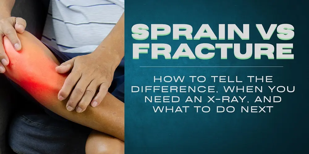
You twisted your ankle on the stairs yesterday. Or you landed on your wrist during a game. Now it’s swollen, it hurts, and one question is stuck in your head: is it just a sprain, or did I break something?
Here is the short answer. A sprain is a stretched or torn ligament, the tough band of tissue connecting bone to bone. A fracture is a crack or break in the bone itself. Both cause pain, swelling, and bruising, which is why symptoms alone cannot reliably tell them apart. Only an X-ray, and sometimes an MRI, can confirm the difference.
That doesn’t mean you’re helpless until you reach a clinic. There are real warning signs that make a fracture more likely, clear situations where you need imaging the same day, and a right way to handle the first 48 hours. This guide walks you through all of it, the way an orthopedic doctor would if you had one sitting next to you.
The 30-Second Difference
Think of your joint as a structure held together by ropes. The bones are the beams, the ligaments are the ropes lashing them together. A sprain means the rope stretched or tore. A fracture means the beam itself cracked.
| Sprain | Fracture | |
| What’s injured | Ligament (soft tissue) | Bone |
| Typical cause | Twisting or rolling a joint | Direct impact, fall, or severe twist |
| Sound at injury | Sometimes a dull “pop” | Sometimes a sharp “crack” |
| Pain centers on | Soft area around the joint | Directly over the bone |
That table is the version most websites stop at. The problem is that real injuries rarely read the table. A bad sprain can hurt more than a small fracture, and some fractures let you walk on them for days. So let’s go deeper, starting with the question your body is already answering.
Where Is Your Pain? A Self-Check Guide
Before any X-ray, doctors do something simple: they press, and they ask where it hurts. You can apply the same logic carefully at home. Work through these four checks.
Check 1: Press gently around the injury, where exactly is the pain?
Pain that is sharpest directly over a bone (the knobby part of your ankle, the edge of your wrist, the shaft of a finger) raises the chance of a fracture. Bone pain tends to be pinpoint: you can place one finger on the worst spot. Pain that spreads through the soft, fleshy area around the joint, and hurts most when you stretch or move it, points more toward a sprain.
Check 2: Can you bear weight?
Try to take four steps (for a leg injury) or grip a light object (for a wrist or hand injury). If you cannot put any weight on the limb, or the pain makes you stop immediately, get medical evaluation; this is one of the criteria doctors themselves use when deciding who needs an X-ray. If you can bear weight but it hurts, that unfortunately rules out nothing: it could still be either injury.
Check 3: Look at the shape.
Compare the injured side with your uninjured side. A joint that looks bent at an unnatural angle, a new lump along a bone, or a limb pointing the wrong direction means fracture until proven otherwise. Go to an emergency department now; don’t wait to see if it improves.
Check 4: Check sensation and color.
Numbness, tingling, pins and needles, or fingers and toes turning pale or bluish suggest a nerve or blood vessel is being compressed, a possible complication of fracture. This also needs emergency care, not a wait-and-see approach.
Important: these checks narrow the possibilities, they cannot confirm a diagnosis. If you finished all four and you’re still unsure, the honest answer is that a doctor with an X-ray machine may be unsure too, until the image is on the screen. Here’s why.
Why Even Doctors Can’t Always Tell Without Imaging
This is the part almost no article tells you, and it’s the most useful thing to understand about your injury.
Pain is a terrible measuring stick. A grade III ankle sprain, a completely torn ligament, is often more painful, more swollen, and slower to heal than a simple stable fracture. Meanwhile, a stress fracture or a hairline crack in a foot bone may let you walk around for a week wondering why it still aches.
Which brings us to the most stubborn myth in sports injuries: “If you can move it, it’s not broken.” This is false, and orthopedic doctors hear it every day. Plenty of people move (and walk on) fractured bones. Movement depends on muscles and tendons, which may be perfectly intact while the bone underneath is cracked. Using that myth as your test is exactly how a small, easily treated fracture becomes a displaced one that needs surgery.
Experienced clinicians know this, which is why they don’t guess either. They examine you, apply decision rules, and confirm with imaging. If a doctor can’t reliably call it from symptoms alone, don’t expect yourself to.
Get a free second opinion from HOSPIDIO’s orthopedic specialists before deciding anything
Sprain vs Fracture: Symptom Comparison
With those limits in mind, here is how the two injuries typically compare, remembering that “typically” is doing a lot of work in this table.
| Symptom | Sprain | Fracture |
| Pain type | Aching, throbbing, worse with movement | Sharp, pinpoint, worse with pressure on the bone |
| Swelling | Often gradual, over hours | Often rapid and significant |
| Bruising | Common, may appear next day | Common, often deep and spreading |
| Sound at injury | Dull pop (ligament tearing) | Crack, grinding with movement afterward |
| Weight-bearing | Painful but often possible | Often impossible, but not always |
| Deformity | Rare | Possible, a strong fracture sign |
| Numbness/tingling | Uncommon | More likely, seek urgent care |
Two rows deserve emphasis. Deformity and numbness are the closest things to definitive home signs; if either is present, stop self-assessing and get seen today. And note the weight-bearing row: “often impossible” is not “always impossible.” Walking does not clear you.
A word about timing, because it worries people: swelling and bruising that appear the next day are common with both injuries and don’t automatically mean anything sinister. Bruising in particular often migrates: blood tracks downward with gravity, so an ankle injury can produce bruising along the foot or toes a day or two later. What matters more than when the swelling arrived is its direction of travel: injuries that are improving each day are reassuring; injuries where pain or swelling is holding steady or worsening after 48 hours have earned an X-ray.
When You Need an X-ray (and When You Might Not)
Get imaging the same day if any of these apply: You can’t bear weight for four steps, there’s a visible deformity, you have numbness or color changes below the injury, pain is directly over a bone, you heard a crack, or the injured person is a child; children’s growing bones fracture in ways that are easy to miss and important to treat.
It may be reasonable to watch and wait 24 to 48 hours if: You can bear weight (painfully but steadily), pain is in the soft tissue rather than on bone, swelling is mild, and things are trending better, not worse. If pain and swelling aren’t clearly improving after 48 hours of proper rest, get an X-ray.
One more thing your urgent-care visit may not mention: A normal first X-ray does not always rule out a fracture. Certain breaks (the scaphoid bone in the wrist is the classic example, along with stress fractures and some subtle ankle fractures) often don’t show on initial images. That’s why a doctor may splint you anyway and repeat the X-ray in 10 to 14 days, or order an MRI. If your X-ray was “clear” but real pain persists for weeks, go back. You’re not imagining it, and asking for follow-up imaging is reasonable, not dramatic.
Age changes the math, too. Children’s bones are softer and still growing, they bend, buckle, and crack through growth plates in patterns that can look almost normal on an X-ray and barely slow the child down, yet can affect how the bone grows if missed. A limping child, or one protecting a limb, should be seen by a pediatric orthopedic specialist rather than watched. At the other end of life, bones weakened by osteoporosis can fracture from surprisingly minor falls, so a lower threshold for imaging is sensible for older adults even when symptoms seem mild.
Where should you go? Emergency department for deformity, numbness, an open wound over the injury, or inability to bear weight at all. Urgent care for same-day X-rays of a stable, walkable injury. Orthopedic clinic for injuries that aren’t emergencies but aren’t improving, and for follow-up of anything the first two find.
Already have an X-ray or MRI? Send your report for a free review by senior orthopedic doctors. You will know within hours whether it needs treatment.
What Happens at the Orthopedic Clinic
Knowing what’s coming makes the visit less intimidating, and helps you give better answers.
First, the history. The doctor will ask exactly how the injury happened: which direction the joint twisted, whether you heard anything, whether you could walk immediately afterward. These details genuinely change the diagnosis, so try to remember them before the visit.
Second, the physical exam. The doctor palpates (presses along specific bones and ligaments in a set order) and tests the joint’s stability and range of motion. This is where clinical decision rules come in: validated checklists (such as the Ottawa ankle rules) that tell doctors which injuries need imaging and which almost certainly don’t. It looks like simple prodding; it’s actually a structured test.
Third, imaging if indicated: usually X-rays first, from two or three angles. If the X-ray is normal but the exam suggests something deeper, such as a torn ligament or a hidden fracture, an MRI or CT may follow.
Finally, the plan. That might be functional treatment for a sprain (bracing and early movement), a boot or cast for a stable fracture, or a surgical consult for an unstable one. You should leave knowing the diagnosis, the healing timeline, and the warning signs that mean “come back sooner.”
Persistent pain is your body telling you the story isn’t over. An orthopedic specialist can finish it properly, with an exam, the right imaging, and a plan. Book a consultation now!!
Quick Guides by Body Part
Ankle. The most common battleground for this question. Rolled ankles are usually sprains, but pain on the bony knobs (the malleoli) or the midfoot suggests fracture. High ankle sprains (pain above the joint line) heal slowly and are often underestimated. Can’t bear weight for four steps? Get an X-ray.
Wrist. Falls onto an outstretched hand injure the wrist more often than any other mechanism. Be especially alert to pain in the “anatomical snuffbox”, the small hollow at the thumb side of your wrist. That’s scaphoid territory, the bone notorious for fracturing invisibly on first X-rays and healing badly when missed.
Finger. “Jammed” fingers are usually ligament injuries, but a finger that looks rotated or crooked when you make a loose fist, or that dislocated and was popped back, needs an X-ray. Buddy-taping a fracture that actually needed alignment can cost you permanent motion.
Foot. The classic trap: stress fractures and small midfoot fractures that let you keep walking. Foot pain that persists beyond a week of rest, especially pinpoint pain on one spot of the foot’s top or outer edge, deserves imaging even if you’re mobile.
Knee. True knee fractures usually follow real trauma and make walking nearly impossible. The more common confusion is ligament tears (ACL, MCL), which can pop, swell fast, and feel unstable. Rapid swelling within an hour of injury is a see-a-doctor sign regardless of cause.
Also Read: Sports Injuries: Types, Symptoms, Treatment and Recovery in India
What to Do Right Now: The First 48 Hours
Whatever the injury turns out to be, the first two days are about protecting it and controlling swelling.
Do: rest the limb and stop the activity that caused the injury. Ice for 15 to 20 minutes at a time, several times a day, with a cloth between ice and skin. Apply a gentle compression bandage (snug, not tight). Elevate the limb above heart level when sitting or lying down. Take simple pain relief if you normally can and follow the label.
Don’t: apply heat, massage the area, or drink alcohol in the first 48 hours; all three increase swelling. Don’t “walk it off” or test the joint repeatedly to see if it still hurts. And don’t tape or splint a visibly deformed limb yourself; keep it still and get help.
If at any point in these 48 hours you develop numbness, spreading severe pain, or skin color changes, stop waiting and seek care immediately.
And once the acute phase passes? For sprains, complete rest beyond the first couple of days can actually slow recovery: gentle, pain-guided movement helps ligaments heal in the right alignment, which is why modern protocols emphasize early motion over long immobilization. But that advice only applies once a fracture has been ruled out. Moving early on an undiagnosed fracture is how small breaks displace. One more reason the X-ray question matters more than the sprain-versus-fracture label itself.
Treatment and Recovery: What the Timelines Really Look Like
Here’s the counterintuitive part: severity doesn’t follow the labels. Recovery does not sort neatly into “sprain = quick” and “fracture = slow.”
A mild (grade I) sprain typically settles in one to three weeks with rest and progressive movement. A moderate (grade II) sprain, a partial tear, takes roughly three to six weeks, often with a brace and physiotherapy. A complete (grade III) tear can take three months or more, occasionally needs surgery, and without proper rehab can leave a permanently unstable, repeatedly re-sprained joint.
A stable, well-aligned fracture, by contrast, usually heals in six to eight weeks in a cast or boot, often with a very predictable course. Displaced or unstable fractures need realignment, sometimes surgical fixation with plates or screws, and run three months or longer including rehabilitation.
Read those numbers again, the worst sprain outlasts the average fracture. This is why “it’s just a sprain” is the most dangerous sentence in this whole topic, and why a proper diagnosis matters even when nothing is broken.
Whichever injury you have, rehabilitation is not optional extra credit, it’s half the treatment. A joint that healed in a cast or brace comes out stiff and weak, and a healed ligament without retraining tends to re-tear. A few weeks of guided physiotherapy is usually the difference between a joint you trust and an ankle that “goes” every few months for years. The goal of treatment isn’t only healing tissue; it’s making sure the joint comes back strong, stable, and trustworthy.
Want a clear treatment and recovery plan? Get a free second opinion from HOSPIDIO’s orthopedic specialists before deciding anything.
The Bottom Line
You can narrow the odds at home (pain on bone versus soft tissue, weight-bearing, shape, sensation) but you cannot confirm a diagnosis, and neither can anyone else without imaging. If your injury isn’t clearly improving after 48 hours, if pain sits directly on a bone, or if any red flag on this page applies, get it looked at.
If your injury is not an emergency but has not improved after 48 hours, an orthopedic consultation is the most direct route to the right diagnosis and treatment. We can help you find and book a qualified orthopedic specialist online.
Note: This article is for educational purposes and is not a substitute for professional medical diagnosis or treatment. If you believe you have a serious injury, seek medical care promptly.
Recent Blogs




Frequently Asked Questions
Yes, sometimes. Stress fractures, small foot fractures, and stable ankle fractures can all allow walking, usually with pain. Being able to walk does not rule out a broken bone. Walking on an undiagnosed fracture can displace it and turn a simple injury into one that needs surgery.
Many patients walk or limp on fractured feet for days or even weeks before getting an X-ray. The rule of thumb: if you cannot take four steps right after the injury, or if pain sits on a specific point of bone and is not improving after 48 hours, get imaging
Go back to a doctor. Some fractures do not appear on the first X-ray, including scaphoid fractures in the wrist, stress fractures, and certain subtle ankle and foot fractures. Persistent pain weeks after a normal X-ray deserves a repeat X-ray, an MRI, or a CT scan.
This situation is far more common than most people realize, and it is one of the most frequent stories patients share online. A first X-ray is a snapshot, not a guarantee. Some cracks only become visible after 10 to 14 days, once the bone around the fracture line begins to change. You are not overreacting by asking for follow-up imaging.
A pop is more often a ligament injury, since ligaments can make a popping sound when they tear. A sharp crack points more toward bone. Neither sound is reliable on its own, because sprains and fractures can each produce either noise, and many serious injuries happen silently.
Pay more attention to what happened next: whether you could bear weight, where the pain settled, and how fast the swelling arrived. A pop followed by rapid swelling and a feeling of instability suggests a significant ligament tear, which can be just as serious as a fracture and also deserves proper evaluation.
Not necessarily. Delayed swelling and bruising are common with both sprains and fractures. Bruising often appears a day or two later and can travel below the injury as blood tracks downward with gravity. The direction matters more: an injury that is improving daily is reassuring; one that is worsening needs an X-ray.
A bruised toe after an ankle injury, for example, often alarms people, but it is usually gravity moving old blood, not a new injury. What should prompt a visit is swelling that keeps increasing after 48 hours, pain that stays fixed over one point of bone, or any numbness or color change.
Yes. A grade III sprain, meaning a completely torn ligament, can be more painful, more swollen, and slower to heal than a stable fracture. Severe sprains can take three months or longer, occasionally need surgery, and can leave lasting joint instability if they are not rehabilitated properly.
A stable fracture in a cast often heals predictably in six to eight weeks. A badly torn ligament has no such schedule. This is why “it’s just a sprain” can be a costly conclusion, and why a proper diagnosis and physiotherapy plan matter even when the X-ray shows no break.
No. This is the most common myth in bone injuries. Movement depends on muscles and tendons, which can work normally over a cracked bone. Many people move, bend, and even use fractured fingers and toes. Pinpoint pain over the bone, rotation, or a crooked appearance matter far more than movement.
A useful check for fingers: make a loose fist and look at your nails. If one finger looks rotated or crosses over its neighbor, that suggests a fracture that needs alignment. Taping a broken finger yourself without ruling this out can cost permanent motion.
Sometimes, yes. Most sprains heal without surgery, but a complete ligament tear that leaves the joint unstable, a sprain that keeps giving way after months of rehabilitation, or certain high-grade injuries in athletes may need surgical repair or reconstruction. This decision usually comes after imaging and a period of guided physiotherapy.
Surgery is the exception, not the rule. The more common mistake is the opposite one: skipping rehabilitation entirely, which leaves the joint weak and prone to repeat sprains. If your joint still feels untrustworthy months after a sprain, see an orthopedic specialist rather than accepting it as normal.
Yes, many can. Small stable fractures are often treated with a walking boot, a splint, or buddy taping to the next finger or toe. What matters is that the bone fragments stay aligned while they heal. That judgment call belongs to a doctor with an X-ray, not to guesswork at home.
Some fractures need more than a cast, including displaced or unstable breaks that require realignment or surgical fixation with plates and screws. So “no cast” can mean either a mild injury or an untreated one. The only way to know which is imaging.
Use the 48-hour rule. If pain and swelling are clearly improving with rest, ice, compression, and elevation, watchful waiting is reasonable. If there is no clear improvement after 48 hours, get an X-ray. If red flags appear at any point, do not wait at all.
Those red flags: inability to take four steps, visible deformity, numbness or tingling, skin turning pale or blue below the injury, an open wound over the injury, pinpoint pain directly on a bone, or an injured child. Any one of these means same-day evaluation.
Go to the ER for deformity, numbness, an open wound, or complete inability to bear weight. Choose urgent care for a same-day X-ray of a stable, walkable injury. See an orthopedic specialist for injuries that are not improving, for follow-up after imaging, and for a recovery and physiotherapy plan.
Dr. Basim Parvez is a licensed physiotherapist and Senior Patient Consultant at HOSPIDIO, holding an MBA in Health Management. With extensive clinical experience and a compassionate approach, he assists patients navigating medical treatments. Dr. Basim also leverages his writing talent to simplify complex healthcare information, empowering patients to make informed decisions and fostering clarity and confidence in their medical journeys.
Sasmita Bal is a Digital Marketing and Content Specialist at HOSPIDIO with expertise in SEO and international healthcare content. She reviews published material to ensure it is optimized for search visibility and relevant to the needs of international patients seeking treatment in India. All content she reviews is authored and clinically approved by the Founder of HOSPIDIO and relevant medical specialists prior to publication.