
Hearing “prominent bronchovascular markings” in your X-ray or CT report can sound alarming, but in most cases, it is a descriptive observation rather than a diagnosis. For some, it reflects temporary changes from an infection; for others, it may hint at chronic lung or heart issues that need monitoring. The best approach is to discuss your report with your doctor, share any ongoing symptoms, and undergo further tests if advised. With the right context, this radiological finding can serve as an early clue, guiding timely intervention and better respiratory health.
In this blog, we will discuss what bronchovascular markings are, why they sometimes appear “prominent”, normal vs. abnormal causes, conditions linked to increased markings, diagnostic significance and when to seek medical advice.
What Are Bronchovascular Markings?
In simple terms, bronchovascular markings are the visible outlines of the lungs’ airways (bronchi) and blood vessels (vascular structures). On a chest X-ray, CT scan or tomography, they appear as thin, branching, web-like lines spread throughout the lung fields. These markings represent the normal structure of the lungs, and everyone has them.
Radiologists examine their thickness, density, and pattern to determine whether they fall within the normal range or appear more pronounced than usual, which may indicate temporary changes from infections, inflammation, fluid buildup, or other conditions that require further evaluation.
A few common reasons include:
- Mild Irritation or InflammationEven something as ordinary as a recent cold or throat infection can cause the airways to become mildly irritated. When the lining of the bronchi swells, they cast a slightly stronger shadow on the X-ray. Allergies can do the same - pollen, dust, or pet dander may trigger temporary inflammation that makes the markings appear more obvious. These changes usually settle on their own once the irritation improves.
- A Bit of Congestion or Extra Blood FlowYour lungs are constantly adjusting to what your body needs. During an infection, fever, or even after intense physical activity, the blood vessels inside the lungs may carry more blood than usual. This increased flow makes them look thicker or more prominent on a scan. It doesn’t always indicate disease, sometimes it’s simply your body responding to stress, exertion, or early congestion that resolves with rest or treatment.
- Fluid or Mucus Around the AirwaysWhen the airways produce extra mucus, whether from a viral illness, allergies, or exposure to irritants, the surrounding tissues can hold more moisture. This can make the walls of the bronchi look sharper or more outlined on imaging. Think of it like tracing a line with a slightly wetter marker; the edges look clearer. Once the mucus clears, the markings often return to their usual appearance.
- Your Natural Body StructureSome people simply have lungs that show more detail. Children, teenagers, and adults who are naturally slim often have less fat and soft tissue around the chest. Because of this, the X-ray beam passes through more easily, making even normal airway and vessel lines appear more defined. In these cases, visible markings may be completely normal for that person.
- Technical FactorsNot all X-rays are identical. Small variations such as how deep a breath you took, the angle of the machine, or the sharpness of the image, can significantly change how the markings appear. A very high-quality or high-contrast X-ray may make normal structures look bolder, while slightly rotated or overexposed films can exaggerate them. Radiologists consider these technical factors before deciding whether a finding is truly significant.
In most cases, this visibility is just a descriptive note for the doctor, not a red flag. It’s one piece of information that needs to be interpreted along with your symptoms, medical history, and any other test results. Many people with more visible markings are completely healthy.
Looking For a Second Opinion From Pulmonologist? Contact the Doctors Below


Normal vs. Abnormal Causes
Normal / Benign Reasons
- Children and young adults: Their lung tissue is thinner, making markings naturally more visible.
- Thin or underweight individuals: Less chest wall fat means lung details show up more clearly.
- Imaging technique: A very sharp or high-resolution X-ray may make markings look exaggerated.
Abnormal / Pathological Reasons
- Infections (acute or chronic bronchitis, pneumonia)
- Pulmonary congestion due to heart problems
- Chronic lung diseases such as COPD, asthma, or interstitial lung disease
- Occupational exposure to dust, fumes, or allergens
- Smoking-related changes that thicken airway walls
Conditions Commonly Linked to Increased Markings
Increased lung markings on a chest X-ray can result from various temporary or chronic conditions affecting the airways, blood vessels, or lung tissue. Below are some common medical causes that can lead to this radiological finding:
1. Respiratory Infection: Viral or bacterial bronchitis, pneumonia, or even post-viral lung inflammation can temporarily increase markings. This often resolves after treatment and healing.
2. Asthma and Allergic Airway Disease: Recurrent airway inflammation can make bronchial walls thicker, showing up as prominent markings. Asthma patients may also show “peribronchial cuffing” (ring-like shadows around bronchi).
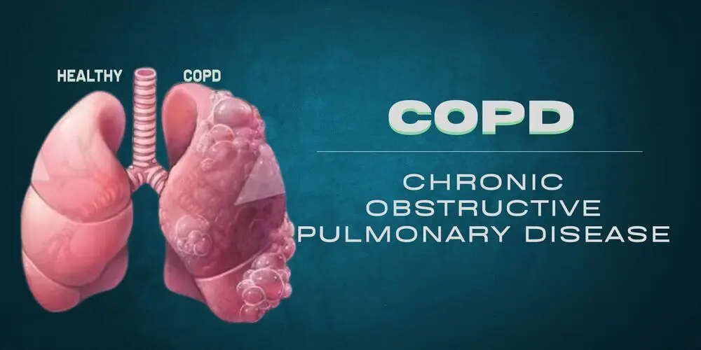
3. Chronic Obstructive Pulmonary Disease (COPD): Long-term smoking or exposure to irritants leads to structural changes in airways and vessels. Radiology often shows coarse, thickened bronchovascular markings, especially in advanced cases.
4. Pulmonary Venous Hypertension / Congestion: In heart failure or valvular heart disease, excess blood backs up into the lungs.This causes vascular markings to become more prominent, especially in the lower lung zones.
5. Interstitial Lung Disease: A group of disorders (like pulmonary fibrosis) that stiffen and scar lung tissue. Early stages may show subtle thickening of markings before progressing to more obvious changes.
6. Environmental or Occupational Lung Injury: Prolonged exposure to coal dust, asbestos, or silica can cause structural lung changes. Markings may become more defined due to inflammation and fibrosis.
Also Read:Lung Transplant in India
Symptoms That May Accompany This Finding
If prominent bronchovascular markings are associated with symptoms, they may indicate an underlying condition. Watch for:
- Persistent cough
- Shortness of breath or wheezing
- Chest pain or heaviness
- Recurrent lung infections
- Fatigue, especially on exertion
- Swelling in ankles (in heart-related causes)
Asymptomatic patients with this finding often don’t need aggressive workup unless other risk factors are present.
What Should You Do If Your X-Ray Report Shows This?
If your chest X-ray report mentions prominent bronchovascular markings, your first step is to consult a pulmonologist or chest physician not to panic. The finding by itself is not a diagnosis; it is a signal that something in your lungs or heart deserves closer attention. Here is a practical checklist:
- Do not ignore the report. Even if you have no symptoms, a follow-up consultation is recommended.
- Ask for a CT scan if your doctor recommends it. A CT scan gives a far more detailed picture than an X-ray.
- Share your full medical history - smoking history, allergies, past respiratory infections, and any heart conditions.
- If you are outside India and want a specialist second opinion at affordable cost, HOSPIDIO can connect you with experienced pulmonologists at accredited hospitals in India within 24–48 hours.
Get a Second Opinion from a Pulmonologist in India
Underlying Conditions & Treatment Costs in India
| Underlying Condition | Common Treatment | Estimated Cost in India |
| Chronic bronchitis / COPD | Pulmonology consultation + bronchodilator therapy | $200 - $800 |
| Congestive heart failure | Cardiac evaluation, medication, possible intervention | $1,000 - $8,000 |
| Pneumonia / respiratory infection | Antibiotic therapy, hospitalisation if needed | $300 - $1,500 |
| Pulmonary hypertension | Specialist cardiology consultation + tests | $500 - $2,500 |
| Asthma / allergic airway disease | Pulmonology management + allergy testing | $150 - $600 |
*All costs are approximate and depend on hospital tier and city. Hospidio helps international patients access JCI and NABH-accredited hospitals at transparent, pre-agreed prices.
Find the Right Pulmonologist or Cardiologist for Your Condition
When Should You Be Concerned?
Not every report mentioning “prominent bronchovascular markings” is worrisome. However, you should seek medical advice if you have:
- Persistent or worsening respiratory symptoms
- History of smoking or long-term pollutant exposure
- Known heart disease, diabetes, or hypertensionFamily history of lung or cardiac conditions
- Abnormal lab tests (e.g., low oxygen, abnormal ECG, high inflammatory markers)
A doctor may recommend additional tests like a chest CT scan, pulmonary function test, or echocardiogram depending on your history.
Can They Be Prevented or Reversed?
The visibility of markings themselves isn’t usually a “disease” but a sign. Preventive strategies focus on lung and heart health:
- Quit smoking: The most effective step for reducing airway changes.
- Manage allergies & asthma: Use prescribed inhalers and avoid triggers.
- Heart health: Control blood pressure, diabetes, and cholesterol to prevent pulmonary congestion
- .Vaccinations: Annual flu shots and pneumonia vaccines reduce infection risk.
- Healthy lifestyle: Exercise, balanced diet, and maintaining healthy body weight support lung capacity.
Why International Patients Choose India for Lung & Chest Treatment
A pulmonologist consultation at a top accredited hospital in India typically costs between $30 - $80 USD as compared to $200 - $400 or more in the UK, US, or Australia. Diagnostic tests like CT scans and pulmonary function tests are similarly priced at 60 - 80% below Western rates, without any compromise on equipment or expertise.
India's leading hospitals including Apollo, Fortis, Max, and Medanta hold JCI or NABH accreditation, the same international quality standards recognised globally. Their pulmonology and cardiology departments handle complex respiratory and cardiac cases from patients across Africa, the Middle East, Southeast Asia, and the Pacific Islands every day.
HOSPIDIO coordinates the entire process for you at no cost. We match you with the right specialist for your condition, assist with the medical visa, arrange hospital appointments, and stay with you through every step from your first consultation to your return home. There are no facilitation fees charged to patients.
Talk to a HOSPIDIO Medical Travel Advisor - Free Consultation
References
- Redcliffe Labs
- Doctors Diagnostic Centre
Read More Blogs


Recent Blogs


FAQs
It refers to airways and blood vessels in the lungs appearing more visible or thicker than usual on a chest X-ray or CT scan. It is a descriptive radiological finding, not a diagnosis. In most cases it points to an underlying condition that requires further evaluation by a pulmonologist or chest physician.
Common causes include respiratory infections such as bronchitis or pneumonia, chronic conditions like asthma or COPD, early congestive heart failure, smoking, and in some cases normal anatomical variation. A follow-up consultation with a specialist is the best way to identify the specific cause in your case.
There is no specific medicine for the markings themselves since they are a sign, not a disease. Treatment targets the underlying cause, antibiotics for infections, inhalers or bronchodilators for asthma, corticosteroids for inflammation, or cardiac medications for heart-related causes. Your doctor will recommend the right approach after diagnosis.
The most effective approach is to treat the root cause. This means quitting smoking if applicable, managing asthma or allergic conditions, following your prescribed cardiac treatment if heart disease is involved, and completing any prescribed course of antibiotics for infections. In many cases the markings reduce significantly once the underlying condition is controlled.
You should consult a doctor promptly if you experience persistent cough, shortness of breath, chest tightness, recurrent respiratory infections, or if you have a known history of heart or lung disease. Even without symptoms, a specialist review is advisable since some conditions that cause these markings such as early heart failure or COPD benefit significantly from early treatment.
Yes. India is one of the most popular destinations for international patients seeking pulmonology and cardiology treatment, offering world-class care at a fraction of the cost compared to the UK, US, or Australia. Hospitals such as Apollo, Fortis, Max, and Medanta are JCI and NABH accredited and have dedicated international patient departments. HOSPIDIO helps patients from across the world access these hospitals with free coordination, medical visa support, and no facilitation fees.
A pulmonologist consultation at a top hospital in India typically costs between $30 - $80 USD. Diagnostic tests such as a CT scan of the chest range from $80 - $200, and pulmonary function tests cost approximately $30 - $60. These prices are 60 - 80% lower than equivalent consultations and tests in the UK, Australia, or the United States. Hospidio can provide a personalised cost estimate based on your specific condition and preferred hospital.
Sasmita is a Marketing Specialist at Hospidio, a leading medical travel company. With expertise in Google Ads, Facebook Ads, and SEO, she plays a pivotal role in driving international leads for healthcare services in India. In addition to her digital marketing prowess, Sasmita is passionate about creating informative and research-based content. She writes extensively about treatment options available in India, the leading hospitals, and the surgeons that provide specialized care. Her blog posts also explore into new medical technologies and breakthroughs in the healthcare field, with the aim of educating international patients on the benefits of traveling to India for medical treatment.
Guneet Bhatia is the Founder of HOSPIDIO and an accomplished content reviewer with extensive experience in medical content development, instructional design, and blogging. Passionate about creating impactful content, she excels in ensuring accuracy and clarity in every piece. Guneet enjoys engaging in meaningful conversations with people from diverse ethnic and cultural backgrounds, enriching her perspective. When she's not working, she cherishes quality time with her family, enjoys good music, and loves brainstorming innovative ideas with her team.