
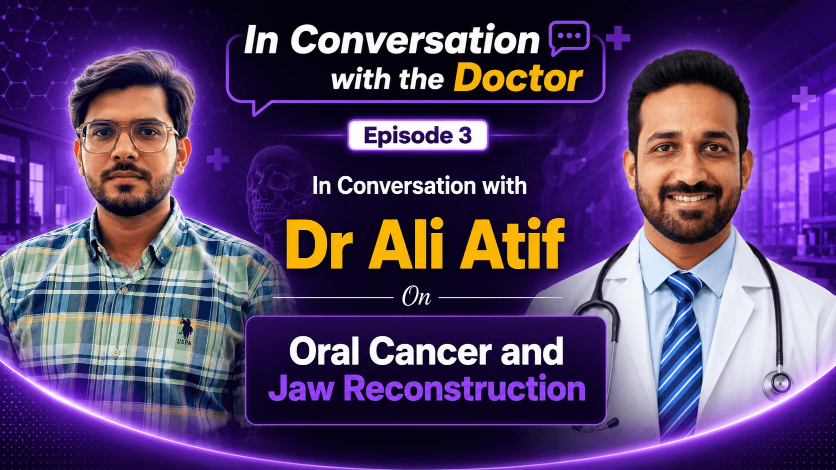
In this Q&A, Dr. Ali Atif, an oral and maxillofacial surgeon in Lucknow with 15 years of experience in head and neck oncology, answers the most common questions patients and families ask about oral cancer, from early warning signs and risk factors to surgery, jaw reconstruction, and what the future holds.
Watch the full conversation:
Q: For someone who has never heard the term "oral cancer," what exactly is it, where can it develop, and why is it considered so serious?
Oral cancer starts inside the oral cavity, which is why it carries that name, but it tends to spread into distant regions of the body as well. It can develop on the tongue, the lip, the floor of the mouth, the cheek, or even the jawbone.
In a healthy person, cells develop and divide in a controlled, normal way. In a cancer patient, cells lose that normalcy and begin multiplying at a very fast, unstoppable rate, forming a tumorous growth. This growth can travel from the oral cavity into the lymph nodes, and from there to the lungs, bones, and other distant sites. Oral cancers are particularly aggressive compared to many other cancers, which is exactly why early diagnosis and awareness matter so much.
Q: Who is most at risk of developing oral cancer? Tobacco, alcohol, and betel nut are often mentioned as major risk factors, what is the single biggest driver?
Tobacco use is by far the biggest risk factor. This includes chewing tobacco, which is very common in India, and smokeless tobacco, which is widely used in African countries.
Alcohol use alongside tobacco has a synergistic effect, increasing risk further. Chewing areca or betel nut can lead to a condition called oral submucous fibrosis, where fibrosis develops around the mouth and reduces mouth opening over time, a precancerous condition that can later progress into cancer. Human papillomavirus (HPV) infection is another contributing factor; biopsies of removed cancerous lesions sometimes show HPV involvement. Across all of these factors, tobacco remains the single biggest driver.
Q: What are the early warning signs of oral cancer that patients often ignore or mistake for something harmless, like a mouth ulcer? How long do patients typically wait before seeing a doctor, and why does that delay matter?
Not every mouth ulcer is oral cancer, so there's no need for alarm over every sore. That said, certain signs warrant attention: an ulcer that persists for many months, white patches inside the mouth, fibrosis that gradually reduces mouth opening, a burning sensation when eating spicy food, or a hard lump in the mouth or neck.
Most patients don't jump to "this might be cancer." They typically see a dentist first for what looks like a simple ulcer, which is exactly why a dentist trained in oral oncology is often the person best positioned to catch it early. The transition from a simple ulcer to oral cancer isn't a one-day event. The body usually shows precancerous signs first, such as leukoplakia, a white patch inside the cheek that cannot be scraped off, or a progressively reduced mouth opening. If something persists and looks suspicious, it's worth seeing a doctor or dentist without delay.
Q: Can you explain what a jaw tumor is, where it grows, and how it differs from oral cancers of soft tissue like the tongue and cheek?
It's important to first separate a cancer, a malignant problem, from a tumor, which is typically a benign, self-limiting growth. A cancer requires complete surgical resolution along with neck dissection to remove infected lymph nodes, followed by reconstruction. A tumor, on the other hand, is a limited growth that carries some risk of eventually developing into cancer, but isn't cancer itself.
Jaw tumors often originate from tooth-related issues, for example, an infected tooth left untreated can develop into a cyst, which over time can progress into a tumor. Ameloblastoma is a well-known example. Some tumors grow outward, expanding and visibly swelling the jaw, while others grow inward and go unnoticed until a tooth loosens or swelling becomes obvious. When treated appropriately, these tumors are self-limiting and can be removed without spreading. Routine dental checkups are important specifically to catch these early.
Read More: Oral Cancer Treatment in India
Q: Ameloblastoma is one of the most talked-about jaw tumors. Why is it so concerning despite being benign, and what happens if it's left untreated?
Ameloblastoma is a rare, slow-growing tumor that usually forms in the lower jaw, in the back teeth region near where wisdom teeth are typically found. It expands internally, resorbing the surrounding bone as it grows, and can spread extensively in multiple directions.
What makes it particularly concerning is its malignant potential. If left untreated, it can develop into cancer over time. This is why diagnosis always starts with a biopsy to rule out cancer, followed by a definitive reconstructive treatment plan. Because it grows so slowly and often goes unnoticed, late diagnosis sometimes means having to remove a significant portion, or even all, of the jaw.
Q: How does an ameloblastoma typically present? At what stage do symptoms like bad breath or swelling start to appear?
Unlike many other conditions, this is largely a painless swelling in its early stages, which is why patients often don't seek care right away. Once pain begins, it usually signals that the tumor is starting to involve the teeth, causing them to loosen. This creates spaces where food can get lodged, and infection can set in alongside the tumor itself, leading to bad breath (halitosis) and red, swollen gums.
This is typically the stage at which patients finally seek treatment, later than ideal, but even at this point, the condition remains treatable if not ignored further.
Q: Can you walk us through what happens during oral cancer surgery? What gets removed, how wide are the surgical margins, and how do surgeons ensure all the cancer is taken out?
Oral cancers are staged using the TNM classification, which accounts for tumor size, the number of lymph nodes involved, and the potential for metastasis, meaning whether the cancer has spread to distant lymph nodes. Surgeons assess this through physical examination, feeling for hardened, indurated margins around the affected area, which indicate cancerous tissue.
When removing the tumor, a safety margin of around 2.5 cm of healthy tissue is taken around it, even if the visible tumor looks small, to minimize the risk of recurrence. This is an aggressive, ablative surgery by necessity, since overly conservative margins significantly increase the chance of the cancer returning.
Alongside removing the primary tumor, a neck dissection is performed to remove lymph nodes at various levels (levels 1 through 5) that may be infected. These are sent for biopsy to determine exactly which levels were involved, which then guides decisions on whether the patient needs radiotherapy, chemotherapy, or both.
Q: If part of a patient's jaw has to be removed, what happens next? Can jaw reconstruction really restore a normal life?
Yes. Modern reconstruction has come a long way. Using digital modeling and 3D-printed jaw structures, surgeons can now recreate a missing portion of the jaw with a high degree of precision. One of the most established techniques is the free fibula graft, where a bone from the patient's own leg (the fibula) is harvested, shaped into a jaw-like structure, and placed in the affected region complete with its own blood supply.
Dental implants can even be placed into this reconstructed bone later, allowing patients to return to a largely normal life, including eating and speaking, after the healing process.
Q: Beyond the free fibula graft, what other reconstruction options are available?
For cheek or soft tissue reconstruction, localized flaps are often used when a free flap isn't cost-effective or medically suitable. Options include the nasolabial flap (using nasal skin to reconstruct the floor of the mouth), neck muscle flaps, and the pectoralis major flap, which uses chest muscle to reconstruct the inner lining of the cheek or provide fuller cheek reconstruction.
For more complex cases, particularly tongue reconstruction after a hemiglossectomy (partial tongue removal), a free radial forearm flap can be used. Remarkably, patients who undergo this kind of reconstruction can often regain near-normal speech, sometimes to the point where it's difficult to tell they've had major surgery at all.
Q: What is the realistic functional and cosmetic outcome after reconstruction?
Outcomes are generally very good, though some tightening or scarring can occur, particularly in patients who also undergo radiation or chemotherapy, both of which affect tissue healing. Laser treatments and other cosmetic interventions are often used afterward to help correct scarring and improve symmetry.
With digital modeling, surgeons can create a 3D-printed model of the patient's skull, match the reconstruction to the healthy contralateral side, and achieve results that are often difficult to distinguish from a patient who hasn't undergone major surgery.
Q: How long does surgery for major jaw resection and reconstruction take, and what does the patient experience in the days and weeks afterward?
Timing depends heavily on the reconstruction method used. Removing the primary tumor along with neck dissection typically takes about 2 to 2.5 hours. Adding a localized flap adds another 1 to 2 hours, bringing the total to roughly 3 to 4 hours for surgery with local flap reconstruction.
Free flap reconstruction takes considerably longer, since it involves connecting blood vessels between the donor site and the oral cavity. Bone-based free flap reconstruction can take 7 to 8 hours or more for complex cases, while soft tissue free flaps, like the radial forearm flap, typically take 5 to 6 hours.
Immediately after surgery, patients are kept in ICU care for the first day, often with a nasal feeding tube and sometimes a tracheostomy to safeguard the airway. By the second or third day, patients are encouraged to walk and regain mobility. Because the inside of the mouth is healing, all feeding happens through the nasal tube for about 7 to 10 days, after which sutures are removed and the patient can begin eating soft or liquid foods orally. Full healing typically takes around 21 days, by which point biopsy results and staging are available to guide any further treatment, such as radiotherapy or chemotherapy. Most patients are discharged around the third day post-surgery, with the first 72 hours being the most critical window.
Q: What is the single most effective thing someone can do to reduce their risk of oral cancer?
Avoiding tobacco, alcohol, and other substance use is the single most impactful step. Safer sex practices also matter, given the role HPV can play. A healthy, balanced diet rich in vegetables, antioxidants, and protein supports overall risk reduction, especially for anyone managing comorbidities like diabetes or hypertension alongside other risk factors.
Routine dental checkups are an underused but highly effective prevention tool. Anyone quitting tobacco after learning about these risks should also make it a habit to stay in regular contact with a dental clinic experienced in oral oncology, since these specialists are best positioned to catch problems at the earliest, most treatable stage.
Q: What excites you most about the future of oral cancer and jaw tumor treatment, robotic surgery, targeted therapies, better reconstructive materials, or AI in surgical planning?
All of the above. Robotics has already made a significant impact on surgical outcomes. HPV vaccines and ongoing research into oral cancer vaccination are promising developments on the prevention side. Stem cell research is opening the door to generating healthier tissue and scaffolds, potentially reducing the need for highly ablative surgery and minimizing morbidity.
Targeted therapies, AI-assisted surgical planning, and continued global research funding are all pushing the field toward less invasive, more precise treatment with better quality-of-life outcomes for patients.
Q: For patients in Africa or the Middle East, where oral cancer and jaw tumor treatment may be unavailable or unaffordable, is traveling to India a realistic option? What should they look for in choosing a center?
Yes, and increasingly so. Many patients from these regions face a lack of specialized infrastructure or costs that simply aren't accessible, even where skilled doctors are present locally. India has become a major destination for this kind of care, with well-established systems for planning every part of the journey, from airport pickup and accommodation to diet and follow-up care.
India is home to a large number of highly experienced cancer specialists, and hospitals here have treated a significant number of patients from Africa and the Middle East, with strong referral networks already in place. Both the medical expertise and the logistics of traveling for treatment have become considerably more accessible and affordable in recent years.
About Dr. Ali Atif
Dr. Ali Atif is an oral and maxillofacial surgeon based in Lucknow, India, with 12 years of experience in head and neck oncology alongside expertise in facial aesthetic and reconstructive surgery. Learn more about his practice and book a consultation.
Considering oral cancer or jaw tumor treatment in India? HOSPIDIO connects international patients with leading specialists and top-accredited hospitals across India, offering transparent pricing, treatment planning, and end-to-end travel support.
Recent Blogs


Dr. Basim Parvez is a licensed physiotherapist and Senior Patient Consultant at HOSPIDIO, holding an MBA in Health Management. With extensive clinical experience and a compassionate approach, he assists patients navigating medical treatments. Dr. Basim also leverages his writing talent to simplify complex healthcare information, empowering patients to make informed decisions and fostering clarity and confidence in their medical journeys.
Guneet Bhatia is the Founder of HOSPIDIO and an accomplished content reviewer with extensive experience in medical content development, instructional design, and blogging. Passionate about creating impactful content, she excels in ensuring accuracy and clarity in every piece. Guneet enjoys engaging in meaningful conversations with people from diverse ethnic and cultural backgrounds, enriching her perspective. When she's not working, she cherishes quality time with her family, enjoys good music, and loves brainstorming innovative ideas with her team.