

It often begins quietly. A dull ache in the groin after a long walk, a stiffness getting out of the car, a twinge that seems to come and go with no obvious pattern. Many patients we speak with are in their thirties or forties, active and otherwise healthy, and the pain gets waved away as a muscle strain or the early signs of arthritis that surely cannot apply to someone their age. An X-ray often comes back reassuringly normal. It is only when the pain refuses to settle, and an MRI is finally ordered, that the real picture emerges: avascular necrosis of the hip.
If you have just received this diagnosis, or you are trying to understand what it means for someone you care about, this guide walks through what avascular necrosis actually is, why it happens, how doctors stage it, and the full range of treatments available today, from watching and waiting through to hip preservation surgery and joint replacement.
What Is Avascular Necrosis of the Hip?
Avascular necrosis, also called osteonecrosis, is the death of bone tissue caused by an interruption to its blood supply. Bone, like every other living tissue in the body, needs a constant flow of blood to stay healthy. When that supply is cut off or significantly reduced, the bone cells in the affected area begin to die. In the hip, this almost always affects the femoral head, the ball shaped top of the thigh bone that sits inside the hip socket.
In the early stages, the bone structure can still hold its shape even as the cells within it are dying, which is exactly why symptoms and early imaging can be misleading. Over time, however, the weakened bone can no longer support the forces passing through it during standing and walking, and the surface of the femoral head begins to collapse. Once that collapse reaches the joint surface, the smooth cartilage covering the bone is damaged too, and a form of secondary arthritis sets in.
It is worth drawing a clear line between avascular necrosis and ordinary osteoarthritis, because the two are often confused. Osteoarthritis is a gradual, mechanical wearing down of cartilage over decades, typically affecting older adults. Avascular necrosis is a vascular event, a genuine interruption of blood supply, and it can affect people in their twenties, thirties and forties who have no other joint problems at all. It is also not unusual for both hips to be affected, sometimes at different stages of progression, particularly when the underlying cause is linked to steroid use or alcohol.
What Causes It
Avascular necrosis has a wide range of possible causes, and they generally fall into two groups: traumatic and non-traumatic.
Traumatic causes are the most straightforward to understand. A hip fracture or hip dislocation can directly damage the blood vessels supplying the femoral head, cutting off its blood supply at the source. This is why avascular necrosis is a recognised, watched-for complication after certain hip injuries, sometimes appearing months or even years after the original trauma has apparently healed.
Non-traumatic causes are more varied, and in many cases several factors overlap. The most commonly cited include long-term or high-dose corticosteroid use, prescribed for conditions ranging from autoimmune disease to organ transplantation, which is one of the strongest known risk factors. Heavy, sustained alcohol use is similarly well established as a contributing factor. Sickle cell disease and other blood disorders that affect circulation or clotting substantially raise the risk, particularly in younger patients. Radiation therapy to the pelvis, decompression sickness in divers, and certain autoimmune conditions are less common but recognised contributors.
In a meaningful proportion of cases, no clear cause is ever identified, and these are described as idiopathic. This is frustrating for patients who understandably want an explanation, but it does not change the approach to treatment, which is guided by the stage and extent of the disease rather than its cause.
A Newer Cause Worth Knowing: Post-COVID and Post-Corticosteroid AVN
In recent years, a distinct and better documented cause has emerged: avascular necrosis linked to corticosteroid treatment given during COVID-19 infection. High-dose steroids such as dexamethasone and methylprednisolone became a standard part of treatment for moderate and severe COVID-19 worldwide, and a growing body of published research has since linked this treatment to a rise in AVN cases, particularly of the hip, in the months and years that followed.
What makes this form of the condition worth understanding on its own is how differently it behaves compared with classic steroid-induced AVN. Traditional steroid-related AVN is generally associated with high cumulative doses, often in the range of two grams of steroid or more, taken over six to twelve months. Post-COVID cases described in recent clinical studies have developed at markedly lower cumulative doses, sometimes under one gram, and over exposure periods as short as two to eight weeks. The disease has also tended to progress more quickly and more aggressively in this group, frequently affecting young, previously healthy adults who had no other risk factors before their COVID-19 illness.
This matters practically for one simple reason: it is easy to overlook the connection. If you had a short course of steroids for COVID-19, or for any other acute illness, some time ago, and are now noticing new or worsening hip or groin pain, it is worth mentioning that steroid course specifically when you see a doctor, even if it feels like ancient history. Recent research following patients with post-COVID AVN over two years found that conservative treatment and core decompression alone tended to offer only temporary improvement in this group, while total hip replacement produced strong, lasting results once the disease had progressed, underlining why an accurate diagnosis and stage, reached sooner rather than later, matters just as much here as with any other cause of AVN.
Symptoms and How the Condition Progresses
In its earliest stage, avascular necrosis can cause no symptoms at all, and is sometimes picked up incidentally on an MRI ordered for an unrelated reason. When symptoms do appear, they typically begin as a deep ache in the groin, sometimes radiating down the front or side of the thigh, or into the buttock. Pain is usually worse with weight bearing activity such as walking or climbing stairs, and eases with rest in the early phases. As the condition progresses, pain can become more constant, present even at rest or disturbing sleep, and range of motion in the hip, particularly internal rotation, becomes noticeably restricted. Many patients develop a limp simply from favouring the joint.
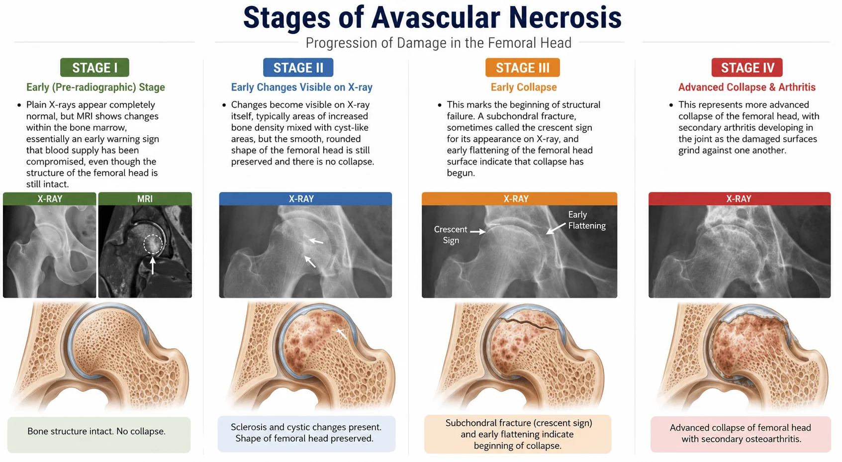
Doctors describe the progression of avascular necrosis using a staging system, most commonly a version of the Ficat and Arlet classification. Understanding the broad stages is genuinely useful for patients, because the stage at diagnosis has a direct bearing on which treatments remain realistic options.
Stage I
Plain X-rays appear completely normal, but MRI shows changes within the bone marrow, essentially an early warning sign that blood supply has been compromised, even though the structure of the femoral head is still intact.
Stage II
Changes become visible on X-ray itself, typically areas of increased bone density mixed with cyst-like areas, but the smooth, rounded shape of the femoral head is still preserved and there is no collapse.
Stage III
This marks the beginning of structural failure. A subchondral fracture, sometimes called the crescent sign for its appearance on X-ray, and early flattening of the femoral head surface indicate that collapse has begun.
Stage IV
This represents more advanced collapse of the femoral head, with secondary arthritis developing in the joint as the damaged surfaces grind against one another.
The practical significance of this staging is straightforward. Stages I and II are described as precollapse, and this is the window in which hip preserving treatments have the best chance of success. Once collapse begins in stage III, and certainly by stage IV, the options narrow considerably, and joint replacement becomes the more reliable path to lasting pain relief.
How It's Diagnosed
Diagnosis begins with a clinical examination, where a doctor will typically check the range of motion in the hip, and look for pain on specific movements, particularly internal rotation. From there, imaging is essential, and this is where avascular necrosis catches many patients out.
A plain X-ray is usually the first test ordered, largely because it is quick, inexpensive and widely available. The difficulty is that X-rays can appear entirely normal in the early stages of the disease, which is precisely when treatment options are widest. A normal X-ray in a patient with persistent hip or groin pain and known risk factors, such as steroid use, heavy alcohol use or sickle cell disease, should not be treated as reassurance that nothing is wrong.
MRI is the gold standard for early detection. It can identify the bone marrow changes associated with avascular necrosis well before any change is visible on X-ray, often showing a characteristic pattern sometimes referred to as a double line sign at the boundary between healthy and affected bone. Where MRI is unavailable or contraindicated, a bone scan can offer useful supporting information, though it is generally less specific.
Treatment Options, From Conservative to Surgical
Treatment for avascular necrosis sits along a spectrum, and where a patient falls on that spectrum depends heavily on the stage of disease at diagnosis, the size and location of the affected area, and individual factors such as age and activity level.
Conservative management
In the very earliest, smallest lesions, doctors may recommend a period of protected weight bearing, using crutches to reduce load through the joint, alongside pain management and physiotherapy for the surrounding muscles. Addressing any underlying cause, tapering corticosteroid doses where medically safe to do so, managing sickle cell disease, or reducing alcohol intake, is an important part of this approach regardless of what other treatment follows. Medications such as bisphosphonates have been studied for their potential to slow progression, though the evidence remains mixed. It is worth being clear eyed about the limits of conservative treatment: on its own, without addressing the underlying vascular problem, many precollapse lesions will continue to progress over time, which is why conservative management is often used as a bridge while a firm diagnosis and stage are confirmed, rather than as a definitive long-term solution for anything beyond the smallest, earliest lesions.
Hip preservation surgery
For patients diagnosed at stage I or stage II, before collapse has occurred, hip preservation techniques aim to save the patient's own joint. Core decompression is the most established of these procedures. A surgeon drills one or more channels into the femoral head to relieve pressure that has built up inside the bone, and to stimulate new blood vessel growth and bone healing in the area. This is often combined with bone grafting, using either the patient's own bone or processed donor bone to provide structural support, and increasingly with the addition of bone marrow aspirate concentrate or other biological agents intended to encourage healing at a cellular level. A related option, osteotomy, involves surgically repositioning the femoral head so that the necrotic segment is rotated away from the main weight bearing surface of the joint. It is a technically demanding procedure, generally reserved for younger patients with a relatively small, well defined area of disease. You can read more broadly about when non-surgical and surgical approaches are typically weighed against each other for hip conditions in our guide on physical therapy versus surgery for hip and knee arthritis.
Hip resurfacing
Where a patient has good remaining bone stock but the joint has progressed beyond the point where preservation techniques are likely to succeed, hip resurfacing offers a middle path. Rather than removing the femoral head entirely, the surgeon reshapes it and caps it with a metal covering, while the socket is also resurfaced. This preserves considerably more of the patient's natural bone than a full replacement, which can be an advantage for younger, physically active patients, though it is not appropriate once significant collapse or damage to the socket itself has occurred.
Total hip replacement
Once the femoral head has collapsed significantly and secondary arthritis has set in, typically stage III or stage IV disease, total hip replacement is generally the most reliable route to lasting pain relief and restored function. Both the femoral head and the hip socket are replaced with prosthetic components. This is a well established, extensively studied procedure with excellent long-term outcomes, and for many patients diagnosed at a later stage, it offers a far more predictable result than attempting to preserve a joint that has already sustained significant structural damage. Typical cost ranges for this procedure can be found in our guides to hip replacement surgery in India and hip replacement surgery in Turkey.
Because bilateral involvement is common, particularly with non-traumatic causes, doctors will often examine and image the opposite hip as well, even if it is currently symptom free.
Which Treatment Fits Which Stage
With four possible treatment paths available, the natural question is how a surgeon actually decides between them for an individual patient. Stage is the starting point, but it is not the only factor. The size of the affected area within the femoral head matters considerably. A small, localised lesion behaves very differently from one that involves a large proportion of the weight bearing surface, even at the same nominal stage. Age and activity level also weigh into the decision, since hip preservation techniques are generally most worthwhile for younger patients who would otherwise face many decades of living with a replaced joint, while an older, less active patient with the same stage of disease may reasonably be steered toward a more definitive procedure from the outset.
This is where the accuracy of staging becomes genuinely important, not just as a technical detail but as something that materially affects the options available. A patient whose disease is still at stage I or early stage II, correctly identified through MRI, still has hip preservation genuinely on the table. The same patient, if that early window is missed and the disease is allowed to progress to collapse before a specialist opinion is sought, may find that preservation surgery is no longer realistically viable, and joint replacement becomes the only remaining path. This is one of the strongest arguments for seeing a specialist with specific experience in hip preservation surgery, rather than a general orthopaedic opinion alone, as early as possible after diagnosis, since not every orthopaedic surgeon routinely performs or offers core decompression, osteotomy or the biological augmentation techniques that go alongside them.
Why It's Worth Getting a Second Opinion on Your Staging
There is one further wrinkle worth knowing about staging, and it rarely gets mentioned outside clinical literature. Studies that have asked multiple doctors to independently stage the same set of X-rays and MRI scans have found real disagreement between them, and even the same doctor reviewing the same images on a different day has sometimes reached a different conclusion. This is not a criticism of any individual clinician. Ficat and ARCO staging both rely on interpreting subtle radiological detail, and reasonable, experienced doctors can read that detail differently, particularly at the borderline between stage II and stage III, which is precisely the boundary that decides whether hip preservation is still realistically on the table.
The practical takeaway is not to distrust your diagnosis, but to recognise that a second opinion on your specific imaging, ideally from a surgeon who performs hip preservation procedures regularly, is a genuinely useful step before deciding on treatment, rather than an unnecessary extra one. This is especially worth doing if your stage has been described as borderline, or if the recommendation you have been given is to proceed straight to joint replacement while you are still relatively young.
Recovery Expectations by Procedure
Core decompression
This is generally the least invasive of the surgical options. Many patients go home the same day or after one night in hospital, and spend the following four to six weeks on crutches with restricted weight bearing to protect the healing bone. Physiotherapy plays an important role in this period, and most patients return to normal daily activity within six to eight weeks, though higher impact activity is usually restricted for longer.
Osteotomy
This involves a longer recovery, since the surgically repositioned bone needs time to heal fully. Protected weight bearing is typically required for eight to twelve weeks, or longer depending on the surgeon's specific protocol and how the healing is progressing on follow up imaging. Physiotherapy is central to regaining strength and full range of motion over this period.
Hip resurfacing
This typically involves a hospital stay of a few days, with walking, supported by a frame or crutches, usually beginning within a day or two of surgery. Structured physiotherapy over the following six to twelve weeks helps rebuild strength and confidence, and most patients return to the majority of normal activities within about three months.
Total hip replacement
This follows a broadly similar early pattern, with a hospital stay of around two to four days depending on the hospital's protocol and the patient's general health, and walking with assistance usually beginning within twenty-four to forty-eight hours. Physiotherapy continues over six to twelve weeks, and full recovery, including a return to most activities, is generally expected within three to six months. Long-term outcomes for hip replacement are extremely well documented and consistently good.
A Candid Note on Hip Replacement in Younger Patients
Total hip replacement is, deservedly, described as a highly reliable and well proven procedure, and for patients diagnosed at a later stage it is often the right answer. It is worth being equally direct, however, about what that recommendation means for a patient in their twenties, thirties or forties, since this is a group avascular necrosis affects disproportionately compared with ordinary osteoarthritis.
Hip implants have a finite working life. Modern implants are durable and outcomes are consistently good, but most are not expected to last fifty or sixty years of an active younger person's remaining lifetime. This means a patient who has a hip replaced at thirty is likely to need at least one, and possibly more, revision surgeries later in life, as the implant wears or loosens over time. Revision hip replacement is a more complex procedure than a first replacement, with a technically harder surgery and typically a longer recovery.
None of this is a reason to avoid replacement when it is genuinely the right treatment, and for many patients with advanced AVN it clearly is. It is simply a reason to make sure that hip preservation options have been properly and honestly explored first, while they are still viable, rather than moving straight to replacement out of convenience or because a surgeon does not personally offer preservation techniques. A frank conversation with your surgeon about your specific stage, your age, and what a realistic long-term plan looks like, including the likelihood of future revision surgery, is worth having before consenting to any procedure.
Living With the Diagnosis: What to Watch For
Not every patient reading this is facing an immediate decision about surgery, and if you have only recently been diagnosed, or you are still at a precollapse stage, there are practical things worth keeping in mind.
Because bilateral involvement is common, particularly where the underlying cause is steroid use or alcohol related, it is worth asking your doctor whether the opposite hip has been properly assessed, even if it currently feels completely normal. A change in symptoms, whether new pain in the treated hip, pain developing in the other hip, or a previously stable hip becoming more painful, is worth reporting promptly rather than waiting to see if it settles, since repeat MRI can confirm whether the disease has progressed and whether the treatment plan needs to change.
If your avascular necrosis has an identifiable underlying cause, continuing to work with the relevant specialist, whether that is tapering corticosteroids where medically appropriate, managing sickle cell disease, or addressing alcohol use, remains part of protecting the joint regardless of what surgical or non-surgical treatment you pursue.
Finally, and perhaps most importantly, the single biggest factor in preserving your treatment options is time. The earlier a precollapse lesion is identified and assessed by a specialist experienced in hip preservation techniques, the wider the range of options genuinely available to you. Once collapse has occurred, some of those options close, not because they were never suitable, but because the disease has moved past the point where they can reliably work.
A Final Word
If you or someone you love has recently been told they have avascular necrosis of the hip, it is entirely natural to feel unsettled by how much there is to take in: staging systems, treatment options, timelines that vary depending on factors that were probably explained quickly in a busy clinic appointment. You do not need to work all of this out alone. If you would like a second opinion on your imaging and stage, or want to understand what treatment, whether hip preservation, resurfacing or replacement, might look like for your specific case in India or Turkey, we are glad to help you think it through. You can share your reports and MRI with our team at hospidio.com/request-opinion for a personalised, no obligation review.
Recent Blogs




Dr. Basim Parvez is a licensed physiotherapist and Senior Patient Consultant at HOSPIDIO, holding an MBA in Health Management. With extensive clinical experience and a compassionate approach, he assists patients navigating medical treatments. Dr. Basim also leverages his writing talent to simplify complex healthcare information, empowering patients to make informed decisions and fostering clarity and confidence in their medical journeys.
Guneet Bhatia is the Founder of HOSPIDIO and an accomplished content reviewer with extensive experience in medical content development, instructional design, and blogging. Passionate about creating impactful content, she excels in ensuring accuracy and clarity in every piece. Guneet enjoys engaging in meaningful conversations with people from diverse ethnic and cultural backgrounds, enriching her perspective. When she's not working, she cherishes quality time with her family, enjoys good music, and loves brainstorming innovative ideas with her team.